
Page 9 - X. Stem cell biology
P. 9
Differentiation and establishment of epithelial-like cells derived from
human induced pluripotent stem cells by dental pulp epithelial stem cells
Mi Jang, Min-Gi Ki, Jihye Yang, Ji-Hye Kim, Dae-Hyun Jeon and Gene Lee
Lab of Molecular Genetics and Dental Research Institute, School of Dentistry, Seoul National University
INTRODUCTION
Tooth development and regeneration essentially require interaction between epithelial and ectodermal mesenchymal
stem cells. Most studies of tooth development have a problem of securing resource of human epithelial stem cells.
Recently, we reported novel epithelial stem cell lines derived from human pluripotent stem cells using Hertwig’s
epithelial root sheath/Epithelial rests of Malassez (HERS/ERM) cell line. To confirm the reported epithelial stem cell
establishment system, we attempted another dental epithelial resource instead of HERS/ERM for differentiation into
the epithelial cell. In this study, human induced pluripotent stem cells (hiPSCs) were cocultured on dental pulp
epithelial stem cells (DPESC) isolated from the deciduous tooth. Differentiated hiPSCs showed the morphological
change as epithelial-like shape. Differentiated epithelial-like cells on DPESC (dEPI-iPSC) were immortalized with
SV40 large T antigen to overcome a rare population. Immortalized dEPI-iPSC cell line had a normal karyotype, and
short tandem repeat (STR) analysis verified that it was derived from hiPSCs. A similar protein expression pattern
was detected in dEPI-iPSC and DPESC.
RESULTS
Figure 1 Figure 2 Figure3
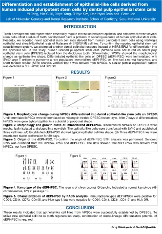
Figure 1. Morphological change during hiPSCs differentiated into dental epithelial-like stem cells on DPESC.
Undifferentiated hiPSCs were differentiated on mitomycin-treated DPESC feeder layer. After 7 days of differentiation,
hiPSCs were grew tightly together in a cuboidal or polygonal shape.
Figure 2. Morphology and growth curve of immortalized dEPI-iPSC. Differentiated hiPSCs on DPESCs were
mechanically isolated and placed in a new dish. The epithelial-like cells were transfected with SV40 and established
three cell lines. (A) Established dEPI-iPSC showed typical epithelial cell-like shape. (B) Three dEPI-iPSC lines were
maintained stable proliferation for 80 days.
Figure 3. Origin of the dEPI-iPSC. To confirm the origin of dEPI-iPSC, STR analysis was performed. Genomic
DNA was extracted from the DPESC, iPSC and dEPI-iPSC. The data showed that dEPI-iPSC was derived from
hiPSCs, not from DPESC.
Figure 4 Figure 5
Figure 4. Karyotype of the dEPI-iPSC. The results of chromosomal G-banding indicated a normal karyotype (46
chromosomes, XY) at passage 10.
Figure 5. Characterization of dEPI-iPSC by FACS analysis. Immunophenotypes dEPI-iPSCs were positive for
CD29, CD44, CD73, CD105, and HLA type I, but were negative for CD90, CD14, CD31, CD117, and HLA-DR.
CONCLUSION
These results indicated that epithelial-like cell lines from hiPSCs were successfully established by DPESCs. To
utilize new epithelial cell line in tooth regeneration study, confirmation of dental-lineage differentiation potential of
dEPI-iPSC is required.
Lab. of Molecular genetics & Stem Cell Differentiation