
Page 5 - X. Stem cell biology
P. 5
Immunomodulatory effect and maintenance of differentiation via
PD1/PDL-1 axis in osteoblasts differentiated from hADMSCs
Seung-Cheol Lee, Min Kyoung Shin and Jung-Suk Sung*
Department of Life Science, Dongguk University-Seoul, Dongguk-ro 32, Ilsandong-gu, Goyang-si,
Gyeonggi-do, South Korea
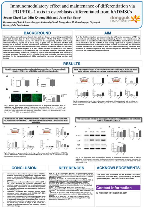
BACKGROUND AIM
Human adipose derived mesenchymal stem cells are known as promising candidates in It is the first investigation on demonstrating the differential expression of PD1 on
stem cell therapy due to their immunomodulatory effects and high potential for cell surfaces elucidating the immunomodulatory function and maintenance of
differentiating into various organs. The study on the adaptability of stem cells using differentiation at osteoblasts differentiated from hADMSCs. Our study may provide
immunomodulatory agents are a basic study to pass over the limitation of stem cell basic information on the PD1/PDL-1 axis having a potential in regulating cellular
therapy and it provides in-depth infrastructure information. PD1 (Programed cell death functions in osteoblasts at the condition with hADMSC. Research for the interaction
protein 1) is known for the immunomodulatory function in immune cells and the anti- between osteoblasts and hADMSCs with their immunomodulatory functions and
tumor activity in various organs. It is also known that MSCs express PD1 and inhibit inhibition of osteoclastogenesis may provide insights in therapeutic strategy to
lymphocyte proliferation by activation of the PD1 pathway. However, functions and the overcome the limitation of stem cell therapy.
molecular mechanism underlying PD1/PDL-1 axis in differentiated cells from hADMSCs
remained unknown. Paracrine activity of cells via various molecular mechanisms at the
condition for the transplantation of MSCs can lead to increased efficacy on stem cell
therapy.
RESULTS
Relative gene expression and protein expression of Programed cell Gene expression levels of pro-inflammatory cytokines in differentiated
death-1 (PD1) on hADMSCs and differentiated cells cells with or without co-culture environments with hADMSCs
Fig. 2. Gene expression levels of pro-inflammatory cytokines in differentiated cells with or without co-
culture environments with hADMSCs (A) Adipocytes with or without hADMSCs (B) Osteoblasts with or
without hADMSCs
Fig. 1. Relative gene expression and protein expression of Programed cell death-1 (PD1) on
hADMSCs and differentiated cells (A) Relative gene expression level of PD1. ***P <0.001 as
compared to ADMSCs (B) Quantitative fluorescent and quantification of fluorescent intensities of PD1
on hADMSCs and differentiated cells (adipocytes, osteoblasts). Green for PD1 and blue for DAPI. Scale
bar = 10 μm, ***P <0.001 as compared to h ADMSCs
Attenuation on gene expression levels of pro-inflammatory cytokines The expression levels of osteogenic markers in osteoblasts co-cultured
by inhibition of PD1/PDL-1 axis in differentiated cells co-cultured with with or without hADMSCs
hADMSCs
Fig. 3. Attenuation on gene expression levels of pro-inflammatory cytokines by inhibition of PD1/PDL-
1 axis in differentiated cells co-cultured with hADMSCs (A) Adipocytes with or without hADMSCs (B)
Osteoblasts with or without hADMSCs
Fig. 4. The expression levels of osteogenic markers in osteoblasts co-cultured with or without
hADMSCs (A) difference in expression of osteogenic markers in osteoblasts and co-cultured osteoblasts (B)
Relative expression of osteogenic markers in osteoblasts and co-cultured osteoblasts, *P <0.05, ***P <0.001
as compared to Os
CONCLUSION REFERENCES ACKNOWLEDGEMENTS
• In this study, we showed that the expression level of PD1 has Baek, S. J., S. K. Kang and J. C. Ra (2011). "In vitro migration capacity This work was supported by the National Research
increased significantly in differentiated osteoblasts when of human adipose tissue-derived mesenchymal stem cells reflects their
expression of receptors for chemokines and growth factors." Exp Mol
compared with that of hADMSCs and the PD1 expression Med 43(10): 596-603. Foundation of Korea (NRF) grant funded by the Korea
level in adipocytes was not significantly differed from that of Bommarito, D., C. Hall, L. S. Taams and V. M. Corrigall (2017). government (MSIT) (NRF-2017R1D1A1B03030983
hADMSCs. "Inflammatory cytokines compromise programmed cell death-1 (PD-1)-
• Gene expression levels of pro-inflammatory cytokines (TNF-α, mediated T cell suppression in inflammatory arthritis through up-
regulation of soluble PD-1." Clin Exp Immunol 188(3): 455-466.
IL-6, IL-1β) decreased in co-cultured osteoblasts with Confalone, E., G. D'Alessio and A. Furia (2010). "IL-6 Induction by
hADMSCs compared with that of non co-cultured osteoblasts. TNFalpha and IL-1beta in an Osteoblast-Like Cell Line." Int J Biomed
Sci 6(2): 135-140.
• Decreased gene expressions of pro-inflammatory cytokines "Mesenchymal Stromal Cell Secretion of Programmed Death-1 Ligands Contact information
in co-cultured osteoblasts were attenuated by inhibition of Davies, L. C., N. Heldring, N. Kadri and K. Le Blanc (2017).
PD1/PD-L1 pathway. Regulates T Cell Mediated Immunosuppression." Stem Cells 35(3): 766-
• Osteoblasts co-cultured with hADMSCs showed higher 776.
osteogenic marker expression levels (RUNX2, ALP, OCN) Lindroos, B., R. Suuronen and S. Miettinen (2011). "The potential of E-mail: hen4118@gmail.com
compared with non co-cultured osteoblasts. adipose stem cells in regenerative medicine." Stem Cell Rev Rep 7(2):
• These results indicate that PD1/PD-L1 axis may played a role 269-291.
Minteer, D., K. G. Marra and J. P. Rubin (2013). "Adipose-derived
in immunosuppressive effect, downregulating TNF-α, IL-6, IL- mesenchymal stem cells: biology and potential applications." Adv
1β expression in osteoblasts differentiated from hADMSCs, Biochem Eng Biotechnol 129: 59-71.
simultaneously maintaining osteogenic marker expressions.
• Our results may provide an insight in the search for a
potential target that can overcome the limitations of stem
cell therapy strategy.