
Page 1 - X. Stem cell biology
P. 1
Exosomes derived from feline adipose tissue
mesenchymal stem cells reduce inflammation factors
Soo-Eun Sung , Kyung-Ku Kang , Joo-Hee Choi, Si-Joon Lee , MinKyoung Sung , Kil-Soo Kim, Min-Soo Seo*
1
Laboratory animal center, Daegu Gyeongbuk Medical Innovation Foundation, Daegu 41061, Republic of Korea
Abstract Results
Adipose tissue derived mesenchymal stem cells (AD-MSCs) release
extracellular vesicles such as exosomes and microparticles.
Particularly, exosomes are formed inward a cell, throughout multi-
vesicular bodies (MVB), so the contents protein, DNA, and RNA are
similar to the parent cells. The exosomes research is rapidly
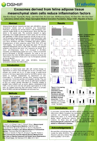
expanding, many studies published in recent years, but the function Figure 1. Characterization
and application of MSC-derived exosomes should emerge as cell- of feline AD-MSCs.
free therapeutics compared with well-known mesenchymal stem cells (A) Isolated feline
therapeutic effects. We isolated exosomes using ultracentrifugation fibroblasts. (B) feline AD-
from feline AD-MSCs and feline fibroblasts cell culture medium. MSC morphology
Characterization of feline exosomes using FACS, DLS, NTA and throughout microscopy
TEM imaging. The exosomes had typical size about 100 nm and (original magnification 100×)
expressed tetraspanins such as CD9, CD81. And then, we analyzed (C) Feline AD-MSC
cytokines and chemokines levels from exosomes by ELISA. As a analyzed stem cells positive
result, anti-inflammatory factor IL-10 increased in AD-MSCs (CD105, CD90, and CD44)
exosomes, pro-inflammatory factors such as IL-1 beta, IL-8 and IFN- and negative (CD14, CD34
gamma decreased in AD-MSC exosomes. Our work is the first and CD45) markers by flow
demonstrated that feline AD-MSCs derived exosomes enhanced the cytometry.
inflammatory suppressive effects and it had the therapeutic potential
of immune disease.
Keywords Mesenchymal stem cells, AD-MSCs, Exosomes,
Extracellular vesicles, Anti-inflammation.
Figure 2. Characterization
Introduction of feline AD-MSC and
fibroblasts exosomes.
Application of mesenchymal stem cells with multiple lineage for (A) Electron micrographs of
regeneration of various tissue damage such as skin, cartilage, bone, isolated exosomes with the
adipose, and muscle and so on. A large number of experimental typical morphology and size.
studies on the tissue regenerative medicine that MSCs secrete many (B) Exosome marker
factors to modulate angiogenesis for regeneration or anti- detection by flow cytometry.
inflammation [1]. Extracellular vesicles (EVs) including exosomes, (C) NTA analysis of
microparticles, membrane particles and apoptotic vesicles. They are exosome particles number
released from various cells under normal of pathological conditions and size distribution.
so they are contain genetic information [2]. Exosome isolation is
possible many biofluids such as serum, plasma, cerebrospinal fluid
(CSF), urine and saliva not only cell culture supernatants [3]. In this
work, we isolated exosomes from cell culture medium to compare Figure 3. Comparing
feline AD-MSCs and dermal fibroblasts derived exosomes. cytokine and
chemokine levels
Methods between feline
fibroblasts derived
[Cell isolation] [Exosome isolation] exosomes and feline
AD-MSCs derived
exosomes. (Y-axis
indicates fluorescence
intensity (a.u), *p-value
< 0.05, **≤0.0001)
Conclusions
In conclusion, isolated feline AD-MSCs derived exosomes reduced
inflammation effects throughout low expression of pro-inflammatory
cytokines and chemokine secretion and high level of anti-
inflammation factor IL-10. Feline AD-MSC and human AD-MSCs
display similar proliferative capacity and immune-suppressive
functions. So these data will support that exosomes have
References immunotherapeutic potentials then it can be used for medicine
related to inflammation diseases.
[1] Han Y, Li X, Zhang Y, Han Y, Chang F, Ding J: Mesenchymal
Stem Cells for Regenerative Medicine. Cells 2019, 8(8).
[2] van der Pol E, Boing AN, Harrison P, Sturk A, Nieuwland R: Acknowledgements
Classification, functions, and clinical relevance of extracellular
vesicles. Pharmacol Rev 2012, 64(3):676-705. We would like to thank laboratory animal resources bank (LAREB) for
[3] Bucan V, Vaslaitis D, Peck CT, Strauss S, Vogt PM, Radtke C: transfer the feline AD-MSCs. This project was by a grant of NLAR
Effect of Exosomes from Rat Adipose-Derived Mesenchymal (National Laboratory Animal Resources) from the Ministry of Food
Stem Cells on Neurite Outgrowth and Sciatic Nerve Regeneration and Drug Safety in 2019-2021.
After Crush Injury. Mol Neurobiol 2019, 56(3):1812-1824.