
Page 9 - N. Metabolism and metabolic diseases
P. 9
Studies of the roles of NADPH oxidase 2 in Gut-liver axis-induced
non-alcoholic fatty liver disease
1
1
Ji Eun Kim , Hye Eun Lee , Yun Soo Bae 1
Department of Life Science, Ewha Womans University, Seoul 03760, Republic of Korea
BACKGROUND AIM
High fat diet (HFD)-induced endotoxemia and dysbiosis allows serum We show Nox2 regulates hepatic injury through endotoxemia and dysbiosis.
lipopolysaccharide (LPS) through increasing permeability of intestinal microbial METHODS
debris and stimulates a low-grade inflammation of various tissues. Recently,
NADPH oxidase 2 (Nox2)-mediated reactive oxygen species (ROS) are involved in To induce NAFLD (Non-Alcoholic fatty liver Diseases) 8-10 weeks old C57BL/6 mice and Nox2
hepatic inflammation. However, no molecular connection between Nox2-mediated gene deficient mice were fed a high fat diet (HFD, 60% of energy from fat, 20% of energy from
protein, and 20% of energy from carbohydrate) for 24 weeks and the control group was fed a
ROS and induction of endotoxemia and dysbiosis has been reported. normal diet (ND, 14% of energy from fat, 27% of energy from protein, 59% of energy from
carbohydrates) for 24 weeks.
RESULTS
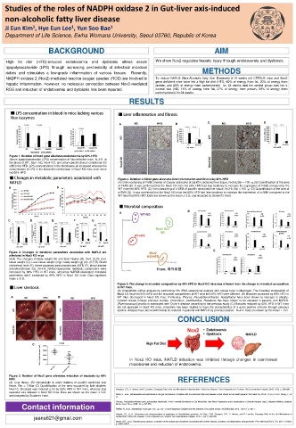
A B C D
Figure 1. Deletion of Nox2 gene alleviates endotoxemia by 60% HFD
Serum lipopolysaccharide (LPS) concentration of Nox-deficient mice. N=4-8. In
the blood of WT, Nox1 KO, Nox4 KO, and colon-specific Duox2 conditional KO
(cKO) fed HFD, LPS concentrations in the blood were all increased whereas the
concentration of LPS in the blood did not increase in Nox2 KO mice even when
fed 60% HFD.
Figure 4. Deletion of Nox2 gene alleviates liver inflammation and fibrosis by 60% HFD
(A) Immunostaining of F4/80 (marker of Cooper astrocyte) of paraffin sectioned liver tissue, N=3-6, Bar = 100 . (B) Quantification of the area
of F4/80 (B). It was confirmed that the Nox2 KO mice fed 60% HFD had less tendency to increase the expression of F4/80 compared to the
A B WT mice fed 60% HFD. (C) Immunostaining of α-SMA of paraffin sectioned liver tissue, N=2-6, Bar = 100 . (D) Quantification of the area of
α-SMA (D). It was confirmed that the Nox2 KO mice fed 60% HFD had less tendency to increase the expression of α-SMA compared to the
WT mice fed 60% HFD. Data are shown as the mean ± S.D. and analyzed by Student’s t-test.
A B
C D E F G
C
Figure 2. Changes in metabolic parameters associated with NAFLD are
alleviated in Nox2 KO mice.
(A,B) The changes of body weight (A) and food intakes (B). N=4. (C,D) Liver
tissue weight (C), Liver tissue weight (mg) / body weight (g) (D). (E,F,G) Blood
cholesterol level (E), blood aspartate aminotransferase (AST) (F), blood alanine
aminotransferase (G). N=4-8. NAFLD-associated metabolic parameters were
increased by 60% HFD in WT mice, wheareas NAFLD-associated metabolic
parameters didn't increased by 60% HFD in Nox2 KO mice. Data represent
mean ± S.D.
Figure 5. The change in microbial composition by 60% HFD in Nox2 KO mice was different from the change in microbial composition
in WT mice.
(A) Unweighted unifrac analysis by performing 16s rRNA sequencing analysis with mouse fecal in Macrogen. The microbial composition of
Nox2 KO mice fed 60% HFD and the microbial compositions of WT mice fed 60% HFD were different. (B) Microbes increased by 60% HFD in
A B WT mice decreased in Nox2 KO mice. Firmicutes, Phocea, Pseudoflavonifractor, Acetatifactor have been shown to increase in obesity-
induced mouse through previous studies. Clostridium, Lactobacillus, Roseburia has been shown to be elevated in patients with NAFLD.
[Ruminococcus] gnavus is associated with Crohn’s disease according to the previous study (C) Microbes reduced by 60% HFD in WT mice
did not decrease in Nox2 KO mice. Kineothrix has been studied to have the characteristics of a gram positive microbe through previous
studies. Alistipes have been confirmed to be reduced in patients with NAFLD by previous studies. N=2-4. Data are shown as the mean ± S.D.
CONCLUSION
C
In Nox2 KO mice, NAFLD induction was inhibited through changes in commensal
microbiome and reduction of endotoxemia.
Figure 3. Deletion of Nox2 gene alleviates induction of steatosis by 60%
HFD REFERENCES
(A) Liver tissue. (B) Hematoxylin & eosin staining of paraffin sectioned liver
tissue. Bar = 100㎛ (C) Quantification of the area occupied by lipid droplets.
N=6-10. Steatosis was induced a lot by 60% HFD in WT mice, whereas less Sharpton, S.R., V. Ajmera, and R. Loomba, Emerging Role of the Gut Microbiome in Nonalcoholic Fatty Liver Disease: From Composition to Function. Clin Gastroenterol Hepatol, 2019. 17(2): p. 296-306.
steatosis was induced in Nox2 KO mice. Data are shown as the mean ± S.D.
and analyzed by Student's t-test. Nobili, V., et al., Bifidobacteria and lactobacilli in the gut microbiome of children with non-alcoholic fatty liver disease: which strains act as health players? Arch Med Sci, 2018. 14(1): p. 81-87. Wang, Y., et
al.,
Phocea, Pseudoflavonifractor and Lactobacillus intestinalis: Three Potential Biomarkers of Gut Microbiota That Affect Progression and Complications of Obesity-Induced Type 2 Diabetes Mellitus. Diabetes
Contact information Metab Syndr Obes, 2020. 13: p. 835-850.
Pfeiffer, N., et al., Acetatifactor muris gen. nov., sp. nov., a novel bacterium isolated from the intestine of an obese mouse. Arch Microbiol, 2012. 194(11): p. 901-7.
Crispim, J.S., et al., Screening and characterization of prophages in Desulfovibrio genomes. Sci Rep, 2018. Sharpton, S.R., V. Ajmera, and R. Loomba, Emerging Role of the Gut Microbiome in
jeana621@gmail.com Nonalcoholic Fatty Liver Disease: From Composition to Function. Clin Gastroenterol Hepatol, 2019. 17(2): p. 296-306.
Henke, M.T., et al., Ruminococcus gnavus, a member of the human gut microbiome associated with Crohn's disease, produces an inflammatory polysaccharide. Proc Natl Acad Sci U S A, 2019. 116(26):
p. 12672-12677.