
Page 17 - N. Metabolism and metabolic diseases
P. 17
The Regulatory function of Hes1 in metabolic disease related macrophage
Da-Hyun kim1,2, Kyung-Hee Chun1,2
1Department of Biochemistry and Molecular Biology, Yonsei University College of Medicine, Seoul, Republic of Korea
2Brain Korea 21 PLUS Project for Medical Science, Yonsei University, Seoul, Republic of Korea
BACKGROUND AIM
Obesity results from a chronic imbalance between caloric intake and energy expenditure that is Adipose tissue macrophages regulated by HES1 in obese
characterized as a low‐grade, chronic inflammatory disease that contributes to metabolic mouse. In this study, we determined the Knockdown of
dysfunction and insulin resistance (IR). Although the molecular basis underpinning this
inflammation is not fully understood, there is consensus that macrophage activation in adipose HES1 in obese mice suppresses obesity, reduces blood
tissue (AT) precedes the development of IR and contributes to a pro‐inflammatory state glucose and CCL2 expression in white fat. Therefore, HES1
Macrophages in obese adipose tissue have been found to populate adipogenic clusters and may be therapeutically targeted to treat obesity and type 2
facilitate angiogenesis and adipogenesis, adipogenic clusters are formed at sites away from
CLSs. HES1 is required for organogenesis and development of several species as a component diabetes.
of the Notch signaling pathway.
METHODS
RNA was isolated using TRIzol® reagent (Invitrogen, Carlsbad, CA, USA), according to the manufacturer's instructions. Reverse transcription-polymerase chain
reaction (RT-PCR) was performed using a reverse transcription system (TOYOBO, Tokyo, Japan) and primers listed in Table 1. PCR was performed using
instructions given in Ex-Taq (TaKaRa, Kyoto, Japan) manual. Real-time PCR was performed using SYBR Premix Ex Taq (Clontech Laboratories, Mountain View, CA,
USA) with ABI instruments (Applied Biosystems Inc, Foster City, CA, USA). All results were normalized by b-actin. All animal experiments were approved by the
Institutional Review Board of the Yonsei University College of Medicine and were performed in specific pathogen-free facilities according to the university’s guidelines
for the Care and Use of Laboratory Animals (2015-0376). 6 weeks old C57BL/6 mouse was purchased from Orientbio. After 1 week of stabilization of mice, fed with a
high fat diet containing 60% fat for 10-12 weeks (12 hours light, 12 hours dark cycle).
RESULTS
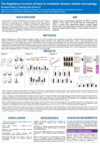
Figure 1 Figure 2 Figure 3 Figure 4
Figure 1: Expression of Notch signaling and HES1 in mouse tissue macrophages. (a) Representative flow cytometry analysis of F4/80 and GFP expression in the SVF of eWAT from normal
chow diet induced LysM cre-GFP mice and high fat diet induced LysM cre-GFP mice. (B-D) Detection of mRNA expression of Notch signaling and HES1 in tissue macrophages from WT
mouse. respectively, B-actin was used as a loading control for RT-PCR analysis.
Figure 2: Expression of Notch signaling and M1,M2 markers in High fat diet induced mouse macrophages using coculture system (a) Illustration of the coculture system composed 3T3-L1
adipocytes and BMDM cells is shown.(B) Detection of mRNA expression of Notch1,3,4 and HES1,2,3,5 in WT mouse BMDM and BMDM coculture with differentiated 3T3-L1 cell from High fat
induced WT mice. respectively, B-actin was used as a loading control for RT-PCR analysis. (B) Detection of mRNA expression of M1 and M2 markes in WT mouse BMDM and BMDM coculture
with differentiated 3T3-L1 cell from High fat induced WT mice. respectively, B-actin was used as a loading control for RT-PCR analysis.
Figure 3: Inhibition of HES1 expression on macrophage improves obesity in High Fat Diet induced mice. Adiposity and weight gain in WT and HES1-LysM cKO mice. (A-B) Body weight of male
WT and HES1 macrophage cKO mice fed a high fat diet were measured over 12 weeks (n = 12 to 15). (C) ) Mice metabolic characteristics. Body weight gain, Body compositions, oxygen
consumption, energy expenditure, Food intake, movement in WT and HES1-LysM cKO HFD mice. For C, *p < 0.05 ; ** p <0.01; and ***p<0.0001.versus WT unpaired t test
Figure 4: Inhibition of HES1 reduced adiposity and improved serum profiles in mice with diet-induced obesity. (A-D)Hematoxylin and eosin (H&E) staining of eWAT, iWAT, Liver and BAT. (B)
Weight of adipocytes and liver. (E) Blood glucose profiles during intraperitoneal glucose tolerance test (GTT)(Left); Blood glucose profiles during intraperitoneal insulin tolerance test
(IPTT)(Right). The results are expressed as the mean ± S.E.M. (n = 4–7/group). (F) Serum levels of the indicated lipid metabolites. For statistical analysis, means and SEM were determined (n =
7 for WT, 7 for cKO [L-E], *, P < 0.05; **, P < 0.01; NS, statistically not significant.
CONCLUSION REFERENCES ACKNOWLEDGEMENTS
. High-fat diet exposure increases Adipose Tissue Djalalinia S, Qorbani M, Peykari N, Kelishadi R. This study was supported by the Bio & Medical Technology
Development Program of the National Research Foundation
Macrophage infiltration. Health impacts of Obesity. Pak J Med Sci of Korea (NRF) funded by the Korean government, MSIP
2015;31:239-42.
HES1 highly express in adipose tissue (NRF-2015M3A9B6073835, NRF-2015M3A9B6073833),
macrophage. Zolkiewska A. ADAM proteases: ligand processing and the NRF grant awarded by theKorean government
and modulation of the Notch pathway. Cell Mol Life (NRF-2019R1A2C289237) to K.H.C.
Adipocyte and macrophage co-culture induced Sci 2008;65:2056-68.
HES1 expression. Bi P, Kuang S. Notch signaling as a novel regulator of Contact information
Downregulation of HES1 reduced lipid metabolism. Trends Endocrinol Metab 2015;26:248-
accumulation in eWAT, iWAT, BAT and Liver. 55. Da-Hyun kim Ph.D. Department of Biochemistry &
Molecular Biology, Yonsei University College of
HES1-LysM cKO mice are resistant to high fat Baek JH, Kim SJ, Kang HG, Lee HW, Kim JH, Hwang Medicine, 50-1 Yonsei-ro, Seodaemun-gu, Seoul
diet–induced obesity and improve glucose KA, et al. Galectin-3 activates PPARgamma and 03722, Republic of Korea Tel.: 82-53-819-1354;
metabolism supports white adipose tissue formation and high-fat
diet-induced obesity. Endocrinology 2015;156:147-56. Fax: 82-2-312-5041; E-mail: dh_kim@dhu.ac.kr