
Page 63 - M. Immunology
P. 63
Enhanced production of IL-23 from dendritic cells by dust mite allergen Der p
upon co-culturing lung epithelial cells
1
Min-Sook Ryu , Jung-In Shin , Ji-Hyun Lee , Perry Ayn Mayson A. Maza , Min-Ho Choi , Jong-Yong Kwak 2,4,5,6
2,3
2
2,3
2,3
1 Department of Allergy, Ajou University School of Medicine, 2 Department of Pharmacology, School of Medicine, Ajou University, 3 Department of Biomedical Sciences, Graduate school, Ajou University, 4 3D
Immune System Imaging Core Center, Ajou University, 5 Immune Network Pioneer Researcg Center, Ajou University, Suwon 16499, South Korea, 6 Nanofaentech Inc. Kimhae 50969, South Korea
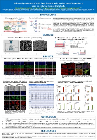
BACKGROUND
Inflammatory mechanism of asthma. The role of IL-23 in pathogenesis of asthma. Airway epithelial cells exposed to activation stimuli, including allergens, viruses, and irritants, release
cytokines that promote dendritic cell (DC) mobilization to draining lymph nodes, where they present
antigens and thereby activate naive CD4 T cells. These T cells then induce B-cell class switching and
maturation into plasma cells, which produce lgE. TH2 cells also migrate into the airway subepithelial
mucosa, where they release inflammatory cytokines such as IL-5 and IL-13, which induce goblet cell
metaplasia and mucus production, and act as a chemokine for eosinophils, mast cells, and basophils.
Unbound lgE secreted by plasma cells binds the FceRI receptor on submucosal mast cells and
basophils and, when crosslinked by an antigen, induces the release of preformed mediators such as
histamine and leukotrienes, as well as the release of inflammatory cytokines. Upon stimulation, mature
dendritic cell (DC) activate naïveT-helper (Th) cells and drive their development into effector Th cells,
such as Th1,Th2, Th17 or regulatory T cells (Treg).Th17cells secrete several effector molecules,
including IL-6, IL-17A/F, IL-23 and trans-forming growth factor-β(TGF-β).
METHODS
Generation of nanofibrous membrane by electrospinning Coculture system with lung epithelial cells and immune
cells in PVA/PCL & PCL/PCL membrane layers
The nanofibers in both membranes were randomly oriented and structurally resembled collagen.
RESULTS
Culture of lung epithelial MLE-12 cells in PVA nanofibrous membranes in the presence of Der P 3D culture of lung epithelial MLE-12 cells in PVA and RGD-PVA
nanofibrous membranes in the presence of Der P
Fig. 1. MLE-12 cells adhered to the nanofibers and were well-distributed throughout the scaffold in PVA nanofibrous Fig. 2. Tight junction formation was lost by Der P treatment and expression
membranes after seeding. The density of phalloidin- and ZO-1-labeled MLE-12 cells exhibiting green and red fluorescences in of beta-actin was increased in MLE-12 cells treated with Der P in RGD-
the membrane significantly decreased 5 d after culturing and der P treatment increased cell detachment from the membrane. PVA.
3D culture of lung epithelial MLE-12 cells in Cytokine production by DCs cultured on culture Increased production of IL-23 and IL-17 in coculture of MLE-12 cells
PVA and YIGSR-PVA nanofibrous membranes dish and nanofibrous membranes and DCs in PVA/PCL & PCL/PCL membrane layers
Fig. 4. The biocompatibility PVA and PCL polymers have
been demonstrated in biological applications. BMDCs cultured
Fig. 3. The adhesion of MLE-12 cells to PVA in PVA and PCL nanofibrous membrane showed inactive
membranes was increased by blending of peptide status in unstimulated condition and fully activated by various Fig. 5. IL-23 and IL-17 production was further increased by coculture of BMDCs, MLE-12
YIGSR of laminin protein which is interact with agonists as in the 2D culture dish. cells and T cells in the presence of Der P in two-layer culture system.
integrin of epithelial cells.
CONCLUSION
1. MLE-12 cells were cultured on PVA membrane for 3 days to form tight junctions between these cells and DCs were cultured on PCL membrane cultured for 4 h for membrane adherence
in each well.
2. After 1 day of treatment with Der p, BMDCs up-regulated expression of MHC-II and CD86 and secreted IL-23 and pro-inflammatory cytokines including TNF-alpha and IL-1beta.
3. Der p failed to induce secretion of IL-23 by MLE-12 cells. Secretion of IL-23 but not TNF-alpha and IL-1beta by Der p-treated DCs was enhanced through co-culturing of MLE-12 cells.
4. The secretion of IL-23 was further enhanced by co-culturing of T cell with DCs and MLE-12 in the presence of Der p. Thus, IL-23 secretion by DCs is enhanced by dust mite allergen-
stimulated lung epithelial cells and further amplified by T cells.
5. DCs are one of the main sources for IL-23 secretion and DC-mediated IL-23 secretion has been considered to be modulated by allergen in lung inflammation.
REFERENCES
Bunte, K. and T. Beikler (2019). "Th17 cells and the IL-23/IL-17 axis in the pathogenesis of periodontitis and immune-mediated inflammatory diseases." International journal of molecular
sciences 20(14): 3394.
Edwards, M. R., et al. (2017). "Addressing unmet needs in understanding asthma mechanisms: From the European Asthma Research and Innovation Partnership (EARIP) Work Package (WP) 2
collaborators." European Respiratory Journal 49(5): 1602448.