
Page 65 - M. Immunology
P. 65
Anti-allergic effect of Catalpa ovata extract on
mast cell-mediated allergic model
a
a
Jae Yeon Lee , Kitae Park , Min Hee Hwang , Hye-Jin Ko , Eun-Kyung Ahn , Yoojin Han and Young-Rak Cho
a*
a
a
a
a
a Bio-Center, Gyeonggi Business & Science Accelerator, 147 Gwanggyo-ro, Yeoungtong-gu, Suwon si, Gyeonggi-do 16229, Republic of Korea
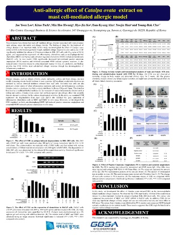
ABSTRACT (A) (B) WBC RBC HGB HCT MCV MCHC PLT
30
6
3
3
Recent studies have shown that mast cell-mediated allergic disorder associated with many diseases 25 Group (10 /μL) (10 /μL) (g/dL) (g/dL) (fL) MCH (pg) (g/dL) (10 /μL) RDW PDW MPV
such asthma, atopic dermatitis and allergic rhinitis. The finding of drugs for the treatment of COE male Body weight(g) Normal 5.7±0.8 9.3±0.5 13.5±0.8 45.6±2.3 49.1±0.7 14.6±0.2 29.7±0.5 610±41.9 13.5±0.3 15.7±0.6 5.0±0.0
500
allergic disease is an important study. In this study, we investigated the effect of Catalpa ovata 20 MPK 4.1±0.5 8.7±0.7 12.7±1.0 42.7±3.4 49.0±0.5 14.6±0.2 29.8±0.4 564±55.8 13.4±0.2 15.8±0.1 5.0±0.1
extract (COE) on the Immunoglobulin E (IgE)-mediated allergic response in vitro and in vivo. COE 1000 5.3±1.9 9.9±0.5 14.3±0.7 48.4±2.4 49.0±0.3 14.5±0.2 29.4±0.3 575±30.7 13.5±0.4 16.3±0.5 5.0±0.1
MPK
significantly inhibited the release of β-hexosaminidase in RBL-2H3 cells with IC value of 94.24 Day 0 1 3 7 14 2000 2.7±0.3 9.4±0.7 13.6±1.2 46.3±3.7 49.4±0.5 14.5±0.2 29.4±0.2 562±52.7 13.7±0.3 16.5±0.2 5.0±0.2
15
50
μg/ml, and suppressed the expression of inflammatory chemokines such as macrophage-derived 25 MPK
RBC
WBC
HCT
HGB
PLT
chemokine (MDC) and thymus and activation regulated chemokine (TARC) in human keratinocyte Group (10 /μL) (10 /μL) (g/dL) (g/dL) MCV (fL) MCH (pg) MCHC (10 /μL) RDW PDW MPV
3
(g/dL)
3
6
(HaCaT) cells. In vivo model, COE significantly decreased IgE-mediated passive cutaneous 20 Normal 3.2±0.5 9.2±0.8 13.9±1.3 46.5±4.0 50.8±0.8 15.2±0.3 30.0±0.2 496±47.3 13.8±0.2 17.2±0.6 5.1±0.3
anaphylaxis (PCA) reaction and inhibited compound 48/80 induced systemic reactions. It also COE female Body weight(g) 500
reduced serum histamine release in a dose dependent manner in mice. In conclusion, our findings 15 MPK 4.2±0.7 9.4±0.1 14.2±0.2 47.5±0.5 50.3±0.3 15.0±0.1 29.8±0.1 492±53.2 13.5±0.2 16.4±0.2 4.9±0.1
1000
provide that COE inhibits mast cell-derived allergic response through the downregulation of MPK 3.9±1.1 9.7±0.4 14.4±0.7 48.5±2.4 50.0±0.3 14.8±0.1 29.7±0.2 524±29.8 13.4±0.1 16.7±0.7 4.8±0.1
10
inflammatory mediators. Day 0 1 3 7 14 2000 3.2±0.8 9.4±0.2 14.1±0.3 47.3±1.1 50.1±0.3 14.9±0.1 29.7±0.2 449±142 13.5±0.3 16.6±0.5 5.0±0.3
MPK
INTRODUCTION Figure. 3. Change in body weight and hematological analysis of male and female ICR mice
during oral administration treated with COE for 14 days. (A) COE was not observed at
mortality, change in body weight and abnormal clinical signs for 2 weeks. (B) The plasma
Allergic diseases, such as allergic rhinitis, atopic dermatitis, asthma and food allergy, are now chemistry analysis did not any hematological toxicities and significant adverse biological effects in
rapidly increasing chronic health problem in most countries. IgE-mediated anaphylactic reactions are all listed clinical chemistry parameters.
mediated by various chemical mediators released from mast cells. Activated mast cells can also (A) (B)
produced a wide variety of other inflammatory mediators and several pro-inflammatory cytokines. 30
Catalpa ovata is a deciduous tree that is widely distributed in Korea, China and Japan. This plant has 25
been used as a traditional herbal medicine for the treatment of various inflammatory diseases such as 20
itching and scabies. Catalpa ovata extract had not been reported anti-allergic effect that scientific Evans blue (μg/ml) 15 * **
data not shown to evidence for the various physiological activities. In this study, we examined COE **
suppressed the degranulation from mast cells and it also inhibited the gene expression of 10 Normal Negative control Positive control
inflammatory chemokines such as MDC and TARC in vitro assay. Moreover, we investigated the 5
COE regulated on both anti-dinitrophenyl(DNP) IgE-induced passive cutaneous anaphylaxis and 0
compound 48/80-induced systemic anaphylaxis in vivo assay. DNP-BSA - + + + + +
COE(mg/kg) 0 0 0 100 200 400
Ketotifen(mg/kg) 0 0 20 0 0 0 100 MPK 200 MPK 400 MPK
RESULTS (C) (D)
500
100 ** **
400 **
80 ** Sample IC 300
Degranulation (%) 60 ** ** COE 94.24 μg/ml Ear thickness (μm) 200 Normal Negative control Positive control
50
40
20 ** Ketotifen 34.76 μg/ml 100
0
0 DNP-BSA - + + + + +
DNP-BSA - + + + + COE(mg/kg) 0 0 0 100 200 400
COE(μg/ml) 0 25 50 100 0 Ketotifen(mg/kg) 0 0 20 0 0 0 100 MPK 200 MPK 400 MPK
Ketotifen(μg/ml) 0 0 0 0 100
(E) (F)
Figure 1 . The effect of COE on antigen-induced degranulation in RBL-2H3 cells. RBL-2H3 150
Compound Blood histamine
5
cells (3X10 per well) were sensitized using 100 ng/ml of mouse monoclonal IgE for 4 hr in 24 Group 48/80 concentration Inhibition
rate(%)
(COE)
well plates. The culture medium was replaced with a PIPES buffer and then treated with various (8 mg/kg) (ng/ml)
90
concentrations of Wheat bran before stimulation with 100 ng/ml of DNP-BSA. The degraulation of No. of mast cell 120 Normal - 6.16±0.89 -
RBL-2H3 cells was determined by the released β-Hexosaminidase activity. Statistical significance 60 Negative control + 25.19±0.93 -
is indicated (*P < 0.05, **P < 0.01 compared with control).
30
Positive control + 6.62±0.49 73.73
(A) 0 100 MPK + 12.09±2.46 51.99
MDC DNP-BSA - + + + + + 200 MPK + 21.77±2.59 13.59
COE(mg/kg) 0 0 0 100 200 400
Ketotifen(mg/kg) 0 0 20 0 0 0 400 MPK + 20.26±3.81 19.57
TARC
Figure. 4. Effect of Passive Cutaneous Anaphylaxis (PCA) reaction and systemic anaphylaxis
GAPDH by COE. The PCA reaction was induced by monoclonal anti-DNP mouse IgE. After 48hr, mice
- + + + + + were injected with antigen(DNP-BSA) in 4% Evans Blue. (A) The values are expressed the amount
IFN-γ,TNF-α(10 ng/ml)
0 0 25 50 100 0
COE(μg/ml) of the dye. (B) The representative pictures of the ears are shown. (C) The amount of extravasated
Dexamethasone(μM) 0 0 0 0 0 100
dye at earskin in mice. (D) The earskin tissues were stained with Toluidine blue O. (E) The change
of mast cells numbers in PCA reaction. (F) The histamine release of COE on compound 48/80
(B) induced systemic anaphylaxis. Statistical significance is indicated (*P < 0.05, **P < 0.01 compared
1.5 3.5
** ** 3 ** ** with control).
MDC mRNA (Densitometric units) 1 ** ** TARC mRNA (Densitometric units) 2.5 ** ** CONCLUSION
2
1.5
0.5
1 In this study, we investigated the effect of Catalpa ovata extract(COE) on the Immunoglobulin
E(IgE)-mediated allergic response. We observed that COE significantly inhibited β-hexosaminidase
0.5 release in a dose-dependent manner in antigen-stimulated RBL-2H3 mast cells. This extract also
0 0 suppressed the expression of MDC and TARC mRNA in HaCaT cells. In addition, COE did not
- + + + + + - + + + + +
IFN-γ,TNF-α(10 ng/ml) show any significant changes of body weight and was not indicated to had any oral toxic effect in
COE(μg/ml) 0 0 25 50 100 0 0 0 25 50 100 0
Dexamethasone(μM) 0 0 0 0 0 100 0 0 0 0 0 100 ICR mice. The extract from Catalpa ovata inhibited the PCA reaction and compound 48/80 induced
systemic anaphylaxis in vivo animal model. These results suggested that COE might be beneficial to
Figure 2. The effect of COE on the expression of chemokines in HaCaT cells. HaCaT cells regulate various allergic reactions.
(1X10 cells) were stimulated with IFN-γ, TNF-α(10 ng/ml) and then treated with COE. (A) Gene
6
expression of chemokines were quantified by RT-PCR. Products were electrophoresed on a 1% ACKNOWLEDGEMENT
agarose gel and staining with ethidium bromide. (B) The relative level of MDC and TARC were
calculated using an image analyzer. Statistical significance is indicated (*P < 0.05, **P < 0.01 This research was supported by Gyeonggi-do, Republic of Korea.
compared with control).