
Page 53 - M. Immunology
P. 53
Immunostimulating activity of a natural plant anthraquinone glycoside, QNG, in murine RAW264.7 macrophages
Hee Jin Kim , Hyeon Jeong Kim , Jisun Lee , Gi Eun Park ,
a
a
a
a
b
a,
Jae Kyung Sohng , and Yong Il Park *
a Department of Biotechnology, The Catholic University of Korea, Bucheon, Gyeonggi-do 14662, Republic of Korea
b Department of Pharmaceutical Engineering, Institute of Biomolecule Reconstruction, Sun Moon University, Asansi, Chungnam 31460, Republic of Korea
* E-mail:yongil382@catholic.ac.kr. Tel:82-2-2164-4512, Fax:82-2-2164-4846
BACKGROUND AIM
Immunostimulation is an important strategy for enhancing the body’s defense This study aimed to assess the potential immunostimulating effects of
systems, especially in the elderly and cancer patients. QNG is a glycoside QNG using RAW 264.7 macrophage. The effects of QNG on pro-
derivative of anthraquinones, present mainly in the roots of a medicinal inflammatory cytokine and chemokine production, iNOS expression, NO
plant Rubia tinctorum. Although QNG has been reported to exhibit various production and cyclin D1 expression, through upregulation of MAPK
bioactivities such as anti-bacterial, anti-cancer and anti-diarrhoeal activities, its signaling were determined.
immunostimulating effect has not been reported.
METHODS
The effect of QNG on secretion of pro-inflammatory cytokine (TNF-α) and chemokine (MIP-1α and RANTES) was assessed by Enzyme-Linked
Immunosorbent Assay (ELISA). The ability of QNG to stimulate NO production was monitored by measuring the intracellular NO concentration using a
confocal miscroscope with a fluorescent NO indicator. The mRNA expression levels of inducible nitric oxide synthase (iNOS) and Interleukin-6 (IL-6) were
measured by RT-PCR. Protein levels of MAPK (ERK and p38) and Cyclin D1 were measured by western blot in RAW 264.7 cells.
RESULTS
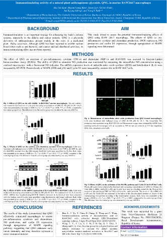
Fig. 1. Effects of QNG on the cell viability in RAW264.7 murine macrophages. The cell viability
was measured after treatment of cells with increasing concentrations of QNG (0-100 μM) for 24 h. Each
value was presented as the means ± SEM (n = 3), *, p < 0.05; **, p < 0.01; ***, p < 0.001, compared to
the control group (Con). The difference from the control group was calculated by one-way ANOVA.
Fig. 4. Measurement of intracellular nitric oxide production from QNG-treated macrophages.
Cells were incubated with indicated doses of QNG (0-100 μM) for 24 h. The intracellular NO
production was monitored using DAF-FM DA and fluorescence images were obtained by LSCM (laser
scanning confocal microscopy) with an excitation at 495 nm and an emission at 515 nm (magnitude
20X). Con, untreated control cells.
Fig. 2. Effects of QNG on the cytokine and chemokine secretion from macrophages. Cells were
incubated with indicated doses of QNG (0-100 μM) for 24 h. The levels of (A) TNF-α, (B) MIP-1α, and
(C) RANTES secreted from the QNG-treated RAW264.7cells were quantified by ELISA. Each value
was presented as the means ± SEM (n = 3), *, p < 0.05; **, p < 0.01; ***, p < 0.001, compared to the
control group (Con). The difference from the control group was calculated by one-way ANOVA.
Fig. 5. Effects of QNG on the activation of the MAPKs pathway and Cyclin D1 in RAW264.7 cells.
Whole cell lysates were collected after treatment with increasing concentrations of QNG for 30 min. (A)
Total ERK (t-ERK), total p38 (t-p38) and β-actin were used as a loading control for the Western blot
Fig 3. Effects of QNG on the mRNA expressions of IL-6 and iNOS in RAW264.7 cells. Cells were analysis of phosphorylated ERK (p-ERK), p38 (p-p38), and Cyclin D1. The levels of (B) p-ERK, (C)
treated with increasing concentrations of QNG (0-100 μM) for 24 h. The expression of (A) IL-6 and (B) p-p38 and (D) Cyclin D1 are expressed as the ratios of the phosphorylated proteins or β-actin to the
iNOS mRNAs was measured using RT-PCR. Relative increases in the levels of each band compared corresponding total protein according to the densitometric analysis. Each value was presented as the
with the loading control β-actin were quantified using densitometry and ImageJ software. Each value means ± SEM (n = 3), *, p < 0.05; **, p < 0.01; ***, p < 0.001, compared to the control group (Con).
was presented as the means ± SEM (n = 3), *, p < 0.05; **, p < 0.01; ***, p < 0.001, compared to the The difference from the control group was calculated by one-way ANOVA.
control group (Con). The difference from the control group was calculated by one-way ANOVA.
CONCLUSION REFERENCES ACKNOWLEDGEMENTS
This work was supported by a grant
The results of this study demonstrated that QNG 1. Han, L., J. Yu, Y. Chen, D. Cheng, X. Wang and C. Wang, from 'Next-Generation BioGreen 21
effectively stimulated macrophages to secrete Immunomodulatory activity of docosahexenoic acid on Program (Project No. PJ0132062020),
pro-inflammatory cytokines and chemokines, RAW264.7 cells activation through GPR120-mediated Rural Development Administration,
NO production, and cyclin D1 expression, 2. signaling pathway, J Agric Food Chem 66(4) (2018) 926-934. Republic of Korea.
Wang, Y. Y., Y. Liu, X. Y. Ni, Z. H. Bai, Q. Y. Chen, Y.
through upregulation of MAPK signaling Zhang and F. G. Gao, Nicotine promotes cell proliferation and
pathway, suggesting that QNG enhances early induces resistance to cisplatin by alpha7 nicotinic Contact information
innate immunity and may therefore represent a acetylcholine receptor-mediated activation in Raw264.7 and E-mail: kcb5077@naver.com
novel immunostimulator. El4 cells, Oncol Rep 31(3) (2014) 1480-1488. Tel: 82-2-2164-4846