
Page 49 - M. Immunology
P. 49
Bee Venom Phospholipase A2 alleviates collagen-induced polyarthritis
by inducing Foxp3+ Treg cell polarization in mice
Gwang-Muk Choi¹, Riwon Hong², Seon-young Park², Hyunsu Bae², Dae-Hyun Hahm¹ , ³*
1 Department of Biomedical Sciences, Graduate School, Kyung Hee University, Seoul, 02447, Republic of Korea
2 Department of Science in Korean Medicine, Graduate School, Kyung Hee University, Seoul, 02447, Republic of Korea
3 Department of Physiology, College of Medicine, Kyung Hee University, Seoul, 02447, Republic of Korea
ABSTRACT AIM
While bee venom (BV) therapy has been widely used for alleviating pain and inflammation in the traditional
medicine, its mechanism is still controversial: constituents-based pharmacological blocking of nociceptive and To investigate that PLA2 can alleviate clinical symptoms of
inflammatory signaling in one side and the BV immunogenicity-based homeopathy-like action in the other side.
In the present study, BV phospholipase A2 (bvPLA2), a BV enzyme catalyzing the release of membrane arthritis and pathological abnormalities of arthritic joints.
arachidonic acids, is suggested to be a novel anti-inflammatory BV mediator stimulating the CD25+ Foxp3+
Treg cell polarization. To verify this hypothesis, anti-arthritic effect of bvPLA2 was evaluated in a mouse model To examine whether the anti-arthritic effect of bvPLA2 is
of rheumatoid arthritis. A collagen-induced experimental rheumatoid arthritis was induced by 2-week-interval
double injections of type II collagen emulsified in complete (1st injection) and incomplete Freund’s adjuvant (2nd) attributed to its action to modulate Treg cell polarization
at the base of the tail. Four days after 2nd injection, bvPLA2 (0.1, 0.5, 1.0 mg/kg) was i.p. injected every two
days for 5 weeks. Arthritic behaviors were significantly alleviated, and histological and micro-CT images of
arthritic joints were highly coincident with their behaviors. Anti-arthritic activity of bvPLA2 became extinct by i.p. To test the hypothesis that injection of bvPLA2 are proposed
injections of 0.25 mg/kg anti-CD25 antibodies (Abs) and 10 μg/kg P60, a peptide inhibitor of Tregs, as regards as a novel therapeutics to treat systemic inflammatory
behavioral assessment. Interestingly, Tregs were completely depleted by the anti-CD25 Abs treatment, not by symptoms of collagen-induced mouse arthritis through the
P60 in flow cytometric analysis of dendritic cells, B-cells and major T cell subsets from spleens. In summary,
bvPLA2 have a significant anti-inflammatory and anti-arthritic activity in rheumatoid arthritis mouse model stimulation of CD4+CD25+ Treg polarization.
through the modulation of development and function of Tregs.
METHODS
s.c. injection of • Two sets of animal studies were performed: one was to examine the anti-arthritic effect of bvPLA2 (Exp. 1) and the other
CII + CFA (1 st ) was about the inhibitory effect by the depletion of Treg cells (Exp. 2)
Histology/µCT/
Flow cytometry
s.c. injection of • 2-week-interval double injections of 100 μg chicken type II collagen (CII) emulsified in complete Freund’s adjuvant (CFA,
CII + IFA (2 nd ) Exp. 1 1 st injection) and incomplete Freund’s adjuvant (IFA, 2 nd injection) to the base of the mouse tail to induced collagen
i.p. injections of PLA2, MTX for 35 days induced arthritis.
Behavioral assessment (twice a week) • Anti-CD25 Abs was i.p. injected two times (day 15 and16) before starting onset of arthritic symptoms (onset stage), and 8
times for 4 weeks after the stage (totally 9 times). Other drugs such as PLA2, P60 (Tregs inhibitor), MTX (positive control)
were injected every 2 days for 32 or 35 days after onset stage. NOR: non-treated naïve group; CIA: collagen-induced
0 50 (day) arthritis group, CIA_PLA2; PLA2 –treated arthritis group; MTX: methotrexate-treated arthritis group; Anti-CD25 Abs: anti-
14 15 16 18 PLA2, P60 ,SCRAMB every 2 days
Exp. 2 i.p. injections of for 32 days mouse CD25 rat Abs-treated arthritis group; P60: peptide 60-treated arthritis group; NOR Abs: normal rat Abs-treated
Anti-CD25 IgG, NOR Abs every 4 days
Anti-CD25 IgG arthritis group; SCRAMB: scrambled peptide-treated arthritis group; PLA2: bee venom phospholipase A2; P60: peptide
P60; MTX: methotrexate; Anti-CD25; Anti-mouse CD25 rat antibodies
RESULTS
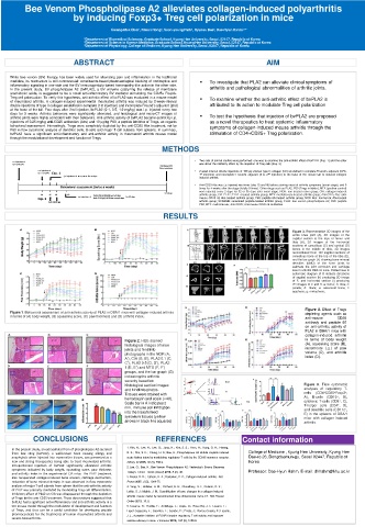
Figure 3. Representative 3D images of the
entire knee joint (A), 2D images of the
sagittal section at the tops of femur and
tibia (B), 2D images of the horizontal
sections of cancellous (C) and cortical (D)
bones in the middle of tibia, 3D images
reconstituted from 100 sagittal sections of
cancellous bone at the top of the tibia (E),
and the bar graph (F) showing bone mineral
densities (BMD) of the knee joints to
estimate the joint corrosion and cartilage
loss in arthritic DBA1/J mice. Dotted lines in
schematic diagram of B indicate directions
of sagittal section (h) producing 3D image
of E, and horizontal section (i) producing
2D images of C and D. a: femur; b: tibia; c:
patella; d: fibula; e: sesamoid bone; f:
epiphysis; g: metaphysis;
Figure 4. Effect of Tregs
Figure 1. Behavioral assessment of anti-arthritic activity of PLA2 in DBA/1 mice with collagen-induced arthritis depleting agents such as
in terms of (A) body weight, (B) squeaking score, (C) paw thickness and (D) arthritis index.. anti-mouse CD25
antibody and peptide 60
on anti-arthritic activity of
PLA2 in DBA/1 mice with
collagen-induced arthritis
Figure 2. H&E-stained in terms of body weight
histological images of knee (A), squeaking score (B),
joints and hindlimb increments ( △ ) of paw
photographs in the NOR (A, volume (C), and arthritis
A’), CIA (B, B’), PLA2 0.1 (C, index (D).
C’), PLA2 0.5 (D, D’), PLA2
1 (E, E’) and MTX (F, F’)
groups, and the bar graph (G)
indicating the arthritic x1000
severity based on FSC H 250 200 150 100 P1 1500 1000 Count
histological section images 105478, 204915 50 0 0 50 100 FSC 150 - A 200 250 x1000 500 0 0 10 2 41.44 CD19 PE 58.56 10 3 - A 10 4 10 5 Figure 5. Flow cytometric
and hindlimb photos. SSC A 250 x1000 200 150 100 P2 4000 3500 3000 2500 Count 2000 analyses of regulatory T-
Tissues were stained with 133398, 116303 50 0 0 50 100 FSC 150 - A 200 250 x1000 1500 1000 500 0 - 10 2 0 10 2 91.69 CD11c APC 8.31 10 3 - Cy7 10 4 - A 10 5 cells (CD4 + CD25 + Foxp3 + ,
hematoxylin and eosin (×40). A 5 10 4 10 CD8 1.64 UR 0.06 A 10 10 5 4 17.20 4.84 A), B-cells (CD19 + , B),
Scale bar in A indicates 2 CD8 APC - 10 10 3 0 3 LL 93.31 - 10 2 0 10 2 10 3 10 4 CD4 4.98 10 5 FoxP3 PerCP 10 0 10 - 10 3 2 2 0 76.71 10 2 10 3 10 4 1.25 10 5 cytotoxic T-cells (CD8 + , C),
mm. immune cell infiltration 10296, 15537 CD4 PE - Cy7 - A 12798, 5481 CD25 FITC - A T-helper cells (CD4 + , D),
and dendritic cells (CD11c + ,
into the transformed E) in the spleens of DBA/1
synovium tissues (yellow mice with collagen-induced
arrows in black line squares) arthritis.
CONCLUSIONS REFERENCES Contact information
In the present study, an extracellular from of phospholipase A2 secreted 1. Kim, H.; Lee, H.; Lee, G.; Jang, H.; Kim, S. S.; Yoon, H.; Kang, G. H.; Hwang,
from bee sting (bvPLA2), a well-known toxin causing allergy and D. S.; Kim, S. K.; Chung, H. S.; Bae, H., Phospholipase A2 inhibits cisplatin-induced College of Medicine , Kyung Hee University, Kyung Hee
anaphylaxis when injected into mammalian tissues, was presented as a acute kidney injury by modulating regulatory T cells by the CD206 mannose receptor. Dae-ro 26, Dongdaemun-gu, Seoul 02447, Republic of
new and strong therapeutics being able to treat rheumatoid arthritis. Kidney Int 2015, 88 (3), 550-9. Korea
Intraperitoneal injection of bvPLA2 significantly alleviated arthritic 2. Lee, G.; Bae, H., Bee Venom Phospholipase A2: Yesterday's Enemy Becomes
symptoms indicated by body weight, squeaking score, paw thickness Professor: Dae-Hyun Hahm, E-mail: dhhahm@khu.ac.kr
and arthritis index in the experimental CIA mice. The PLA2 treatment Today's Friend. Toxins (Basel) 2016, 8 (2), 48.
also recuperated collagen-induced bone erosion, cartilage destruction, 3. Brand, D. D.; Latham, K. A.; Rosloniec, E. F., Collagen-induced arthritis. Nat
reduction of bone mineral density. It was observed in flow cytometric Protoc 2007, 2 (5), 1269-75.
analysis of major T-cell subsets from spleen that the anti-arthritic activity 4. Yang, S.; Hollister, A. M.; Orchard, E. A.; Chaudhery, S. I.; Ostanin, D. V.;
of PLA2 was primarily mediated by modulating Treg cell differentiation. Lokitz, S. J.; Mathis, J. M., Quantification of bone changes in a collagen-induced
Inhibitory effect of PLA2 on CIA was disappeared through the depletion
of Tregs by the anti-CD25 treatment. These observations suggested that arthritis mouse model by reconstructed three dimensional micro-CT. Biol Proced
bvPLA2 had a significant anti-inflammatory and anti-arthritic activity in a Online 2013, 15, 8.
CIA mouse model through the modulation of development and function 5. Casares, N.; Rudilla, F.; Arribillaga, L.; Llopiz, D.; Riezu-Boj, J. I.; Lozano, T.;
of Tregs, and thus can be a useful candidate for developing peptide Lopez-Sagaseta, J.; Guembe, L.; Sarobe, P.; Prieto, J.; Borras-Cuesta, F.; Lasarte,
pharmaceuticals for the treatments of human rheumatoid arthritis and J. J., A peptide inhibitor of FOXP3 impairs regulatory T cell activity and improves
severe osteoarthritis.
vaccine efficacy in mice. J Immunol 2010, 185 (9), 5150-9.