
Page 45 - M. Immunology
P. 45
A Novel Peptide from Spider Venom, Lycotoxin-Pa4a, Exhibits Antibacterial
and Anti-inflammatory Activities
1*
1
2
1
Min Kyoung Shin , In-Wook Hwang , Yunkyung Kim , Seung-Tae Kim , and Jung-Suk Sung
1
1 Department of Life Science, Dongguk University-Seoul, Goyang 10326, Republic of Korea
2 Life and Environment Research Institute, Konkuk University, Seoul 05029, Republic of Korea
BACKGROUND AIM
The emergence of drug-resistant bacteria has become a global issue, increasing the In this study, the transcriptome of an indigenous spider in Korea, Pardosa astrigera (P.
demand for a new source of antibiotics. Antimicrobial peptides (AMPs) are found in a astrigera), were analyzed for the identification of a novel AMP based on comparative
wide range of organisms where it may exhibit multiple functionalities such as analysis of homology and structural characteristics with known toxin peptides. The
immunomodulatory and anticancer effects along with its primary antibacterial activity. potential of utilizing toxin peptide and RNA transcripts for drug design was suggested
AMPs have drawn attention as next-generation antibiotics due to its high selectivity by the investigation of antibacterial activity and immunomodulatory effect. The
and lower side effects. The venom of spider is a rich source of bioactive components, functionality of the toxin component can bring insight into the mode of action of which
as it is used for both predation and defense. the venom acts.
METHODS
Sample preparation – Venom glands of P. astrigera were separated for the extraction of total RNA. Transcriptome library was conducted by subsequent RNA sequencing using
the NGS technique.
Antibacterial activity assay – Colony-forming unit assay was performed using gram-negative (E. coli, P. aeruginosa) and gram-positive bacteria (B. cereus, , S. aureus) to
determine antibacterial activity. Relative colony formation was measured by counting colonies on each plate.
Membrane permeability test – Permeabilization of bacterial outer membrane and cytoplasmic membrane was determined by using dyes NPN and DiSC 3 (5), respectively.
Fluorescence intensity was measured after treatment of the peptide and compared with that of melittin.
NO assay & cell viability assay – Murine macrophage RAW 264.7 were treated with several concentrations of the peptide. Each of supernatant and cells were subjected for NO
measurement and viability test.
RT-qPCR & western blot analysis – After treatment of the peptide with or without LPS, total RNA or protein was extracted from the cells. Gene expression of inflammatory
mediators were measured by RT-qPCR, and change in activity of MAPK pathway was detected via immunoblotting.
RESULTS
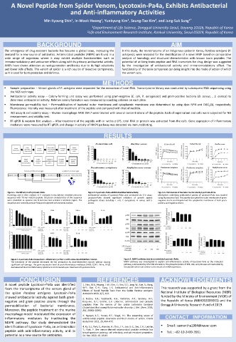
Figure 1. Identification of Lycotoxin-Pa4a. Figure 2. Lycotoxin-Pa4a exhibited antibacterial activity. Figure 3. Permeation of bacterial membrane by Lycotoxin-Pa4a.
Homology and in silico analysis of P. astrigera transcriptome revealed precursor Antibacterial activity of Lycotoxin-Pa4a was conducted via CFU assay. Mechanism underlying antibacterial activity of Lycotoxin-Pa4a was studied
sequence with 75-mer mature peptide. Structural characteristics of the sequence Lycotoxin-Pa4a showed significant inhibition of growth against using fluorescence dye. The peptide disrupted the outer membrane of gram-
were predicted as cysteine-rich N-terminal and α-helical C-terminal region. The pathogenic strains including E. coli, P. aeruginosa, B. cereus, and S. negative strains and depolarized the cytoplasmic membrane of both gram-
sequence was named Lycotoxin-Pa4a and subjected to functional analysis. aureus. positive and negative strains.
Figure 4. Lycotoxin-Pa4a showed anti-inflammatory effect on LPS-stimulated RAW264.7 model. Figure 5. MAPK pathway was inactivated via Lycotoxin-Pa4a.
The treatment of the peptide decreased the NO production by dose-dependent manner without causing MAPK pathway was investigated to explain anti-inflammatory activity of Lycotoxin-Pa4a as the molecular
significant cell damage. The gene expression level of pro-inflammatory mediators (iNOS, COX2, TNF-α, IL-1β) pathway is stimulated by LPS-TLR4 interaction. The phosphorylation of ERK, JNK, and p38 were all inactivated by
decreased while anti-inflammatory cytokine IL-10 increased upon treatment of Lycotoxin-Pa4a. the peptide which was similar to the results of mRNA expression.
CONCLUSION REFERENCES ACKNOWLEDGEMENTS
A novel peptide Lycotoxin-Pa4a was identified 1. Shin, M.K.; Hwang, I.-W.; Kim, Y.; Kim, S.T.; Jang, W.; Lee, S.; Bang,
from the transcriptome of the venom gland of W.Y.; Bae, C.-H.; Sung, J.-S. Antibacterial and Anti-Inflammatory This research was supported by a grant from the
Effects of Novel Peptide Toxin from the Spider Pardosa astrigera.
the spider Pardosa astrigera. Lycotoxin-Pa4a Antibiotics 2020, 9(7), 422. National Institute of Biological Resources (NIBR)
showed antibacterial activity against both gram- funded by the Ministry of Environment (MOE) of
2. Kozlov, S.A.; Vassilevski, A.A.; Feofanov, A.V.; Surovoy, A.Y.;
negative and gram-positive strains through the Karpunin, D.Y.; Grishin, E.V. Latarcins, antimicrobial and cytolytic the Republic of Korea (NIBR202009201) and the
permeabilization of bacterial membranes. peptides from the venom of the spider Lachesana tarabaevi Dongguk University Research Fund of 2019.
(Zodariidae) that exemplify biomolecular diversity. J. Biol. Chem. 2006,
Moreover, the peptide treatment on the murine 281, 20983–20992.
macrophage model modulated the expression of 3. Nguyen, L.T.; Haney, E.F.; Vogel, H.J. The expanding scope of
inflammatory mediators by inactivating the antimicrobial peptide structures and their modes of action. Trends CONTACT INFORMATION
MAPK pathway. Our study demonstrated the Biotechnol. 2011, 29, 464–472.
identification of Lycotoxin-Pa4a, an antimicrobial 4. Ko, S.J.; Park, E.; Asandei, A.; Choi, J.-Y.; Lee, S.-C.; Seo, C.H.; Luchian, Email : samantha1994@naver.com
peptide with anti-inflammatory activity, and its T.; Park, Y. Bee venom-derived antimicrobial peptide melectin has Tel. : +82-10-2499-3561
broad-spectrum potency, cell selectivity, and salt-resistant properties.
potential as a new source for antibiotics. Sci. Rep. 2020, 10, 10145.