
Page 41 - M. Immunology
P. 41
AXL receptor tyrosine kinase-induced autophagy ameliorates acute liver injury by
suppressing NLRP3 inflammasome activation in mice.
Si-Won Park and Taehoon Chun
Department of Biotechnology, College of Life Sciences and Biotechnology,
Korea University, Seoul, 02841, Republic of Korea
BACKGROUND • Autophagy is a homeostatic degradative process which eliminates damaged
• Liver disease is often induced by severe hepatic inflammation which causes liver damage organelles or turns over cytoplasmic constituents through lysosomal compartments in
eukaryotic cells.
and fibrosis. In the progress of hepatic inflammation, macrophages serve as sentinels
which initiate immune responses and mediate wound healing and metabolic functions. AIM
Therefore, it is important to regulate the activity of macrophages for a therapeutic strategy
to ameliorate severe hepatic inflammation. In this study, we explored the function of individual TAM family members during
• The TAM family of RTKs expressed in macrophages downregulates inflammatory autophagy and investigated their contribution in hepatic inflammatory responses.
responses by facilitating the clearance of apoptotic bodies and supplying signals of
inflammation inhibition.
METHODS
Measurement of autophagy induction Cells were treated with various concentrations of recombinant GAS6, CQ and/or serum deprivation for 24 h. Also, various combinations of neutralizing
antibodies or inhibitors were added to block autophagy induction after GAS6 treatment. After treatment, autophagy induction in each cell was measured via immunoblotting and confocal
microscopy. Also, Atg5, Becn1 and Map1lc3b mRNA levels were measured by quantitative reverse-transcriptase polymerase chain reaction (qRT-PCR).
Western blot analyses Cells were washed 3 times with PBS, centrifuged, and lysed with RIPA lysis solution containing 200 mg/ml of phenylmethylsulfonyl fluoride, phosphatase inhibitor
cocktail and protease inhibitor cocktail. The cell lysates were subsequently resolved by 12% SDS-polyacrylamide gel electrophoresis, transferred onto Immobilon P membranes, and
immunoblotted with appropriate antibodies. Immunoreactive bands were visualized using an ECL solution. To determine the MAP1LC3B-I to MAP1LC3B-II ratio, immunoreactive bands were
quantified using ImageJ software version 1.43u. Then, the values of MAP1LC3BI to MAP1LC3B-II ratio were relative to the control value of the experiment. To quantify the relative expression of
other proteins, immunoreactive bands were normalized to ACTB levels.
Confocal microscopy To observe autophagy induction, mCherry-EGFP-MAP1LC3B expressing P388D1 cells and J774 cells were treated with various combinations of recombinant GAS6,
neutralizing antibody and/or inhibitors. The cells were then washed 3 times with PBS and fixed with 4% paraformaldehyde (Sigma, P6148) in PBS at room temperature for 10 min. After washing
3 times with PBS, cells were stained with a mounting solution containing DAPI(Vector Laboratories, H-1200) and imaged by confocal microscopy on an LSM 5 EXCITER (Carl-Zeiss,
Oberkochen, Germany).
RESULTS
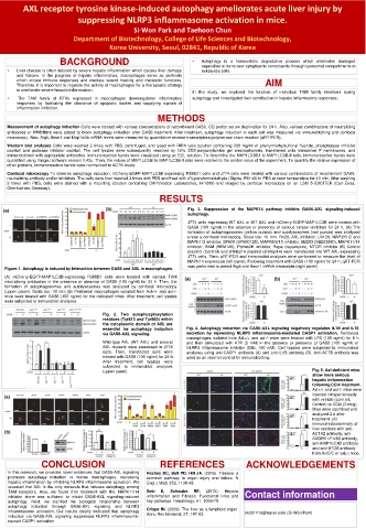
(a) (b) Fig. 3. Suppression of the MAPK14 pathway inhibits GAS6-AXL signaling-induced
autophagy.
J774 cells expressing WT AXL or WT AXL and mCherry-EGFP-MAP1LC3B were treated with
GAS6 (100 ng/ml) in the absence or presence of various kinase inhibitors for 24 h. (A) The
formation of autophagosomes (yellow puncta) and autolysosomes (red puncta) was analyzed
under a confocal microscopy. Scale bar: 10 mm. R428, AXL inhibitor; U0126, MAP2K1/2 and
MAPK1/3 inhibitor; SP600 (SP600125), MAPK8/9/10 inhibitor; SB203 (SB203580), MAPK11/14
inhibitor; BKM (BKM120), PtdIns3K inhibitor; Rapa (rapamycin), MTOR inhibitor (B) Control
plasmid (Control) and shMap14 plasmid (shMap14) were transfected into WT AXL-expressing
J774 cells. Then, qRT-PCR and immunoblot analyses were performed to measure the level of
MAPK14 expression (left panel). Following treatment with GAS6 (100 ng/ml) for 24 h, qRT-PCR
was performed to detect Atg5 and Becn1 mRNA transcripts (right panel).
Figure 1. Autophagy is induced by Interaction between GAS6 and AXL in macrophages.
(A) mCherry-EGFP-MAP1LC3B-expressing P388D1 cells were treated with various TAM (a) (b)
neutralizing antibodies in the presence or absence of GAS6 (100 ng/ml) for 24 h. Then, the
formation of autophagosomes and autolysosomes was analyzed by confocal microscopy
(upper panel). Scale bar: 10 mm (B) Peritoneal macrophages isolated from Axl+/+ and axl-/-
mice were treated with GAS6 (100 ng/ml) for the indicated times. After treatment, cell lysates
were subjected to immunoblot analyses.
Fig. 2. Two autophosphorylation
residues (Tyr815 and Tyr860) within
the cytoplasmic domain of AXL are
essential for autophagy induction Fig. 4. Autophagy induction via GAS6-AXL signaling negatively regulates IL1B and IL18
via GAS6-AXL signaling. secretion by repressing NLRP3 inflammasome-mediated CASP1 activation. Peritoneal
macrophages isolated from Axl+/+ and axl-/- mice were treated with LPS (100 ng/ml) for 6 h
Wild-type AXL (WT AXL) and several and then stimulated with ATP (5 mM) in the absence or presence of GAS6 (100 ng/ml) or
AXL mutants were expressed in J774 NLRP3 inflammasome inhibitor (Glib, 100 mM). Cell lysates were subjected to immunoblot
cells. Then, transfected cells were analyses using anti-CASP1 antibody (A) and anti-IL1B antibody (B). Anti-ACTB antibody was
treated with GAS6 (100 ng/ml) for 24 h. used as an internal control for immunoblotting.
After treatment, cell lysates were
subjected to immunoblot analyses
(upper panel). Fig. 5. Axl-deficient mice
show more serious
hepatic inflammation
following CCl4 treatment.
Axl+/+ and axl-/- mice were
(a) injected intraperitoneally
with vehicle (corn oil,
Control) or CCl4 (2 ml/g).
Mice were sacrificed and
analyzed 2 d after
treatment. (A)
Immunohistochemistry of
liver sections with anti-
(b) ACTA2 antibody, anti-
ADGRE1/F4/80 antibody,
anti-MAP1LC3B antibody
and anti-ATG5 antibody
from AxlC/C or axl¡/¡ mice.
CONCLUSION REFERENCES ACKNOWLEDGEMENTS
In this research, we provides novel evidences that GAS6-AXL signaling Rockey DC, Bell PD, Hill JA. (2015). Fibrosis- a
promotes autophagy induction in murine macrophages, repressing common pathway to organ injury and failure. N
hepatic inflammation by inhibiting NLRP3 inflammasome activation. We Engl J Med, 372, 1138-49.
revealed that AXL is the only molecule that induces autophagy among
(2015).
Hepatic
Seki
E,
Schwabe
RF.
TAM receptors. Also, we found that treatment with the MAPK11/14 inflammation and Fibrosis: Functional links and Contact information
inhibitor alone was sufficient to inhibit GAS6-AXL signaling-induced
autophagy. Next, we clarified the biological relationship between key pathways. Hepatology, 61, 1066-79.
autophagy induction through GAS6-AXL signaling and NLRP3
inflammasome activation. Our results clearly indicated that autophagy Crispe IN. (2009). The liver as a lymphoid organ. kkd01119@naver.com (Si-Won Park)
induction via GAS6-AXL signaling suppresses NLRP3 inflammasome- Annu Rev Immunol, 27, 147-63.
caused CASP1 activation.