
Page 39 - M. Immunology
P. 39
Phc2 Regulates the Migration of Hematopoietic Stem and Progenitor Cell
by Suppressing Vcam1 Expression From Bone Marrow Niche
Jun Yang, Taehoon Chun
Department of Biotechnology, College of Life Sciences and Biotechnology,
Korea University, Seoul, 02841, Republic of Korea
BACKGROUND AIM
● Hematopoietic stem and progenitor cells (HSPCs) sustain the hematopoietic system by ● In this study, we demonstrate that Phc2, a component of the canonical PRC1, regulates
continuously replenishing hematocytes (Wright et.al.,2001). Postnatally, most HSPCs engraft HSPC mobilization through the supression of Vcam1 expression in BMSCs. Therefore,
and reside in the BM niche, but a small fraction of these cells is mobilized and migrate into the Phc2 deficiency causes a severe HSPC mobilization defect via the derepression of
peripheral blood (PB) to reconstitute the hematopoietic system under specific signaling cues Vcam1 in BMSCs, and the pharmacological inhibition of VCAM-1 in BMSCs significantly
(Bhattacharya et.al.,2006). reverses the symptoms of Phc2-deficient mice. These data demonstrate the critical cell-
● Polycomb group (PcG) proteins function as transcriptional repressors of target genes by extrinsic role of Phc2 in controlling HSPC mobilization and provide the first evidence of
modulating histone methylation (Piunti and Shilatifard, 2016). epigenetic control over HSPC mobilization.
METHODS
● Experimental mice and phenotypic analysis
Phc2 +/− (Phc2 heterozygote) mice with a C57BL/6 background were used in this study. C57BL/6 and B6 CD45.1 mice were obtained from The Jackson Laboratory. WT and KO mice were bred
from Phc2 +/− mice. All animals received proper care in accordance with the National Institutes of Health Guide for the Care and Use of Laboratory Animals. The study protocol was approved by
the Institutional Animal Care and Use Committee of Korea University.
● Cell cycle analysis and measurement of apoptosis
BM-resident LSK cells from 8-week-old mice were isolated and incubated in 70% ethanol overnight at 4 °C. After washing with PBS, cells were stained with propidium iodide (Sigma-Aldrich
P4170) for cell cycle analysis. To measure apoptosis, LSK cells were stained with 7-AAD and Annexin V-FITC. After washing with PBS, stained cells were analyzed by flow cytometry.
● CFU assay
Mononuclear cells isolated from the BM, PB, and spleen were plated onto Methocult GF M3434 (StemCell Technologies 03444) in 35 mm cell culture dishes. Cells were then incubated at 37 °C
and 5% CO 2 . After 7 days of incubation, type and number of colonies were determined.
● LSK transplantation
LSK cells (1 × 10 5 ) isolated from 8-week-old donor mice were injected intravenously into lethally irradiated (10 Gy) 8-week-old recipient mice. Twelve weeks after the LSK transfer, recipient mice
were sacrificed and analyzed for reconstitution of donor immune cells. Donor cells and recipient cells were discriminated by flow cytometry using anti-CD45.2 (104)-PerCP-Cy5.5 Ab (BD 552950,
dilution 1:100) for congenic strain (CD45.1) discrimination. For serial competitive LSK repopulation assays 58 , LSK cells (5 × 10 4 ) isolated from WT or KO mice (CD45.2) were mixed with an equal
number of LSK cells isolated from competitor mice (WT in CD45.1). The cell mixture was then intravenously injected into lethally irradiated (10 Gy) 8-week-old recipient mice (WT in CD45.1).
Secondary transplantation was performed at 12 weeks after primary engraftment. LSK cells (1 × 10 5 ) harvested from primary transplants were intravenously injected into lethally irradiated (10 Gy)
8-week-old recipient mice (WT in CD45.1). The ratio of CD45.1 to CD45.2 positive cells in the BM, PB, thymus, and spleen of recipient mice was measured by flow cytometry.
RESULTS
(A) (B) (C)
(A)
(B)
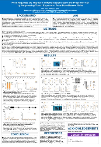
Fig. 2. Impaired recruitment of Phc2 KO HSPCs from the BM into the periphery.
(A) Comparison of WBC counts between WT and KO mice on day 5 after G-CSF treatment. n = 5. (B)
Representative image of spleens from WT and KO mice (left) and absolute numbers of splenocytes (right) for
WT and KO mice on day 5 after G-CSF treatment. n = 5. (C) CFU-C in the BM (femur), PB, and spleen (SP) of
WT and KO mice on day 5 after G-CSF treatment. n =5.
(A) (B)
(C)
(C) (D) (E)
Fig. 1. Downregulation of Phc2 leads to immune cell deficiency
without intrinsic defects in HSPCs.
(A) Absolute number of immune cells in the BM, thymus (Thy), Fig. 3. Phc2 suppresses VCAM-1 expression in BMSCs.
spleen (SP), and liver of WT or KO mice. n = 6. (B) Relative ratio of (A) mRNA expression patterns of genes related to HSPC mobilization in BM niches from WT and KO mice
BM-resident hematopoietic lineage cells between WT and KO mice. were accessed by qRT-PCR. n = 3. (B) Lysates from WT and KO BMSCs were immunoblotted to analyze
n =5. (C) Cell cycle status of BM-resident LSK cells from WT and VCAM-1 expression. n = 3. (C) MFI of VCAM-1 expression in BMSCs from WT and KO mice analyzed by flow
KO BM. n = 4. (D) Percentage of apoptosis for BM-resident LSK cytometry. n = 3 Endo, endothelial cells; OB, osteoblasts; MSC, mesenchymal stem cells.
cells from WT and KO mice. n = 3. (E) BM-resident clonogenic
progenitors from WT and KO mice were assessed by CFU assays.
n = 5. (A) (B) (C)
(A)
(B)
Fig. 5. Blocking the interaction between VCAM-1 and VLA-4 rescues the defect involving regimen-
induced HSPC mobilization in KO mice. (A-C) G-CSF-induced HSPC mobilization assays with anti-VCAM-1
Ab. n = 5 per group. (A) WBC counts from experimental animals until day 5 after G-CSF and Ab treatment. (B)
Representative image of spleens (left) and absolute number of splenocytes from experimental animals on day 5
after G-CSF and Ab treatment (right). (C) CFU-C in the BM (femur), PB, and spleen (SP) from experimental
animals on day 5 after G-CSF and Ab treatment
Fig. 4. Phc2 binds to the Vcam1 gene locus and suppresses which in turn led to a systemic immunodeficiency. stem cell engraftment of rare niches corrects severe
Vcam1 gene expression through histone modifications. Consistent with these results, treatment with a lymphoid deficiencies without host conditioning. J. Exp. Med.
neutralizing Ab against VCAM-1 in Phc2-deficient
(A) Schematic representation of the Vcam1 mice restored HSPC mobilization from the BM into 203, 73–85
gene locus. The locations of primers #1 to #7 and exons are the periphery. These data establish a role for the
indicated. (B) ChIP was performed with WT and KO BM cell lysates epigenetic regulation of VCAM-1 in regulating ACKNOWLEDGEMENTS
using anti-Phc2 Ab (Phc2) or a control Ab (Control). DNA recovered HSPC mobilization, as a key downstream mediator
from ChIP was then analyzed by qPCR. n= 5. of Phc2 functions. None. No funding to declare
CONCLUSION REFERENCES Contact information
Wright, D. E., Wagers, A. J., Gulati, A. P.,
● In this study, we demonstrated that Phc2 binds to the Vcam1 locus Johnson, F. L. & Weissman, I. L. (2001).
to repress its transcription by enhancing H3K27me3 and H2AK119ub Physiological migration of hematopoietic stem and
in BMSCs. In the absence of Phc2, the increased Vcam1 expression in progenitor cells. Science 294, 1933–1936 Jun Yang
BMSCs strengthened the interaction between HSPCs and BMSCs, Bhattacharya, D., Rossi, D. J., Bryder, D. & E-mail : yeejes860@gmail.com
compromising timely HSPC mobilization from the BM into the periphery, Weissman, I. L. (2006). Purified hematopoietic