
Page 55 - M. Immunology
P. 55
Overexpression of p190RhoGEF in macrophage enhances the formation of atherosclerotic plaques
in mouse aorta
1
Eun Bi Lee, Jong Ran Lee 2
¹Department of Bioinspired Science, The Graduate School, Ewha Womans University, Seoul 03760, South Korea,
²Department of Life Science, College of Natural Sciences, Ewha Womans University, Seoul 03760, South Korea
0000000 AIM
To study whether changes in p190RhoGEF expression affect the formation of mouse aortic atherosclerotic plaques,
ApoE mouse was crossed with p190RhoGEF transgenic (TG) mouse and atherosclerotic plaque formation in aorta was
-/-
monitored up to 30 weeks of age.
BACKGROUND
p190RhoGEF was first identified in neuronal cells, where it was shown to be specific for RhoA and its activation regulates
neuronal morphology. Our laboratory has identified p190RhoGEF as one of the molecules, expression of which was
increased after CD40 stimulation in B cells. Previous works in our laboratory showed that overexpression of
p190RhoGEF affects B cell maturation and differentiation. In addition, our studies also showed that p190RhoGEF
overexpression affects negatively several functions of dendritic cells and macrophages in response to
lipopolysaccharides (LPS).
METHODS
-/-
Animals, macrophage preparation, and serum collection. ApoE mice and p190RhoGEF transgenic (TG) mice were cross-bred to
-/-
generate TG/ApoE mice. Cells harvested from the peritoneal cavity were cultured overnight and attached cells (macrophage) were
harvested using a scraper and used for experiments. Blood was collected from the artery and centrifuged. Sera were collected and
used for the measurement of cytokines.
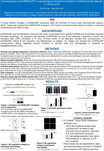
Plaque formation detection. Aorta was perfused through heart with PBS and remaining plaques were fixed with 4%
paraformaldehyde (PFA). Fixed aorta was stained with 0.2% Oil Red O (Sigma) solution. After microscopic photographing, the area of
plaques was detected using Image J and presented by the percentage of total area of aorta.
Cytokine measurement. Ready-SET-Go cytokine ELISA sets were used according to the manufacturer’s instruction. Serum samples
were added to each well of 96-well flat-bottomed ELISA plates that were prepared for the assay. At the end of the assay, the plates
were read at 450 nm using a microplate reader (Molecular Devices, Spectra MAX).
Flow cytometry. Cells were blocked with 3% Bovine Serum Albumin (BSA) solution and stained with antibodies (PerCP-anti-F4/80,
+
+
+
FITC-anti-CD11c, and PE-anti-CD11b). M1 macrophage population (CD11b F4/80 CD11c ) was analysed.
Reactive oxygen species (ROS) measurement. Cells were treated with lipopolysaccharide (LPS; 1 µg/ml) for 30 minutes, followed
by DCF-DA (5 mM) treatment for 15 minutes. Production of ROS was measured using a microscope (Nicon ECLIPSE Ts2R, 20X).
RESULTS
Figure 1. Construct of p190RhoGEF transgene
and its expression.
A) A schematic map of the p190RhoGEF Figure 2. Oil Red O staining in mouse
transgene construct. B) mRNA transcripts of aorta.
p190RhoGEF in macrophage from each animal. *, p < 0.05; mean±S.D., n=11 Figure 3. Serum cytokine level.
**, p < 0.05; mean±S.D., n=5
CONCLUSION
∙In TG/ApoE mouse model compared to
-/-
WT/ApoE mice,
- Aortic atherosclerotic plaques were
increased up to 30 weeks of age.
- The level of serum cytokine, especially
IL-6, was significantly increased.
- M1 macrophage population was higher.
- ROS production in response to LPS
was increased.
Figure 5. M1 population ∙These results may be responsible for rapid
Figure 4. M1 population analysis in analysis in peritoneal
peritoneal macrophage. macrophage. atherosclerotic plaque formation in TG/ApoE -/-
*, p < 0.05; mean±S.D., n=6 mice compared to WT/ApoE control mice.
*, p < 0.05; mean±S.D., n=5