
Page 57 - I. Chemical biology and drug discovery
P. 57
Suppression of angiogenesis by PD in human umbilical
vein endothelial cells
Hyun-Ji Park and Shin-Hyung Park *
1
1
1 Department of Pathology, College of Korean Medicine, Dong-eui University, Busan, 614-052, Korea.
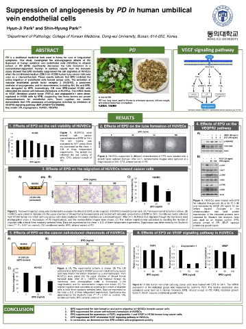
PD is a traditional medicinal herb used in Korea for cure of lung-related
symptoms. This study investigated the anti-angiogenic effects of PD.
Exposure of human umbilical vein endothelial cells (HUVECs) to ethanol
extract of PD (EPD) significantly decreased the tube formation in a
concentration-dependent manner. In addition, results from the transwell
assay showed that EPD markedly suppressed the cell migration of HUVECs
when the conditioned medium (CM) from H1299 human lung cancer cells was
used as a chemoattractant. These results indicate that EPD inhibited the
active migration of endothelial cells toward cancer cells. The activation of
vascular endothelial growth factor receptor 2 (VEGFR2), a prominent
mediator of angiogenesis, and its downstreams, including Akt, Src and ERK,
was abrogated by EPD. Interestingly, CM from EPD-treated H1299 cells
attenuated the cancer cell-induced chemotaxis of HUVECs. The mRNA levels
of VEGF, fibroblast growth factor (FGF)-2, and angiopoietin-1 were down-
regulated in H1299 cells by EPD, suggesting that these factors are potent - A root of PD
chemoattractants for endothelial cells. Taken together, our results - PD has long been used in Korea to eliminate sputum, relieve cough,
demonstrate that EPD possesses anti-angiogenic activities by inhibition of and reduce bronchus contraction.
VEGFR2 signaling pathway (NRF-2019R1F1A1059588). - 祛痰降氣, 宣散風熱
Key words: PD, angiogenesis, HUVEC, VEGFR2
https://www.researchgate.net/figure/VEGF-signaling-pathway-involved-in-angiogenesis_fig1_324776501
Figure 1. HUVECs were
treated with various
concentrations of EPD for 24 h.
The cell viability was
evaluated by MTT assay. Data
are expressed as the mean ±
S.D. of three independent
experiments. The dotted line
indicates the cell viability of Figure 2. HUVECs suspended in different concentrations of EPD were seeded onto a
90%. EPD, ethanol extract of growth factor-reduced Matrigel. After 24 h, representative images were captured at a
PD.
magnification of 50X. EPD, ethanol extract of PD.
Figure 4. HUVECs were treated with EPD
for indicated time periods (A) or for 12 h (B)
and stimulated by VEGF (20 ng/ml) 10 min
before harvest. Changes in the
Figure 3. Transwell migration assay was conducted to evaluate the effects of EPD on the migration of HUVECs toward cancer cells. (A) The experimental scheme is shown. (B) phosphorylation and total protein
HUVECs were plated in triplicate into the upper chamber of 24-well format transwell plate and treated with indicated concentration of EPD for 24 h. Conditioned media collected expressions of the indicated proteins were
from H1299 human non-small cell lung cancer cells were loaded on the down chambers as a chemoattractant. After 24 h, HUVECs that migrated through the membrane were assessed by Western blot analysis. Actin
photographed under a microscope (×100 magnification), and the representative images were shown. (C) The relative migration was calculated by counting the number of was used as an internal control. EPD,
migrated cells at least three randomly selected fields. Data are expressed as the mean ± S.D. of three independent experiments. Significance was determined by the Student's ethanol extract of PD; VEGF, vascular
t-test (*** P < 0.001 vs. control). CM, conditioned media; EPD, ethanol extract of PD. endothelial growth factor.
Figure 5. (A) The experimental scheme is shown. (B) The CM
collected from EPD-treated H1299 human non-small cell lung cancer
cells were filled in the bottom chambers as a chemoattractant. Then
HUVECs were plated into the upper chamber of 24-well format
transwell plate. After 24 h, HUVECs that migrated through the
membrane were photographed under a microscope (×100
magnification), and the representative images were shown. (C) The Figure 6. H1299 human non-small cell lung cancer cells were treated with EPD for 24 h. The mRNA
relative migration was calculated by counting the number of migrated expression of the indicated genes was measured by real-time PCR. The relative expression was
cells at least three randomly selected fields. Data are expressed as calculated using actin as a internal reference. EPD, ethanol extract of PD; FGF2, fibroblast growth
the mean ± S.D. of three independent experiments. Significance was factor 2; VEGF, vascular endothelial growth factor.
determined by the Student's t-test (*** P < 0.001 vs. control). CM,
conditioned media; EPD, ethanol extract of PD.
1. EPD suppressed the tube formation and active migration of HUVECs towards cancer cells.
2. EPD suppressed the cancer cell-induced chemotaxis of HUVECs.
3. EPD suppressed the expression of FGF2, angiopoietin-1 and VEGF in H1299 human lung cancer cells.
4. EPD suppressed VEGF-stimulated VEGF signaling pathway in HUVECs.
5. In conclusion, we demonstrate that EPD exhibits anti-angiogenesis activity.