
Page 55 - I. Chemical biology and drug discovery
P. 55
Hexane fraction of Adenophora triphylla var. japonica triggers
apoptosis by inhibiting STAT3 and Src in human non-small cell
lung cancer cells
Hyun-Ji Park and Shin-Hyung Park 1*
1
1 Department of Pathology, College of Korean Medicine, Dong-eui University, Busan, 47227, Korea.
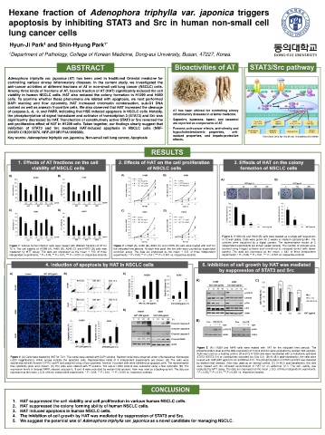
Adenophora triphylla var. japonica (AT) has been used in traditional Oriental medicine for
controlling various airway inflammatory diseases. In the current study, we investigated the
anti-cancer activities of different fractions of AT in non-small cell lung cancer (NSCLC) cells.
Among three kinds of fractions of AT, haxane fraction of AT (HAT) significantly reduced the cell
viability in human NSCLC cells. HAT also reduced the colony formation in H1299 and H460
cells. To examine whether these phenomena are related with apoptosis, we next performed
DAPI staining and flow cytometry. HAT increased chromatin condensation, sub-G1 DNA
content as well as annexin V-positive cells. We also observed that HAT increased the cleavage
of caspase-3, -8, -9, and PARP, indicating that HSE induced apoptosis in NSCLC cells. Notably, - AT has been utilized for controlling airway
the phosphorylation of signal transducer and activator of transcription 3 (STAT3) and Src was inflammatory diseases in oriental medicine.
significantly decreased by HAT. Transfection of constitutively active STAT3 or Src reversed the - Saponins, lupenone, lupeol, and taraxerol
anti-proliferative effect of HAT in H1299 cells. Taken together, our findings clearly suggest that are reported as components of AT.
inhibition of STAT3 and Src mediated HAT-induced apoptosis in NSCLC cells (NRF- - Possess anti-cancer effects, anti-obesity and
2016R1C1B2015076, NRF-2019R1F1A1059588). hypocholesterolaemic properties, anti-
oxidant properties, and hepato-protective
Key words: Adenophora triphylla var. japonica, Non-small cell lung cancer, Apoptosis Front Oncol. 2012 Apr 10;2:30. doi: 10.3389/fonc.2012.00030.
effects.
Figure 3. H1299 (A) and H460 (B) cells were seeded as a single-cell suspension
in 12-well plates. Cells were grown for 2 weeks in medium containing HAT. The
colonies were visualized by a digital camera. The representative results of 3
Figure 1. Various human NSCLC cells were treated with different fractions of AT for Figure 2. H1299 (A), H460 (B), A549 (C) and H1975 (D) cells were treated with HAT for independent experiments are shown (upper panels). The number of colonies were
72 h. The cell viability of H1299 (A), H460 (B), A549 (C) and H1975 (D) cells was the indicated time periods. At each time point, the live cells were counted by trypan blue counted using ImageJ software and normalized to untreated control cells (lower
measured by MTT assay. The data are expressed as the mean ± S.D. of three exclusion assay. The data are expressed as the mean ± S.D. of three independent panels). The data are expressed as the mean ± S.D. of three independent
independent experiments. * P < 0.05, ** P < 0.01, *** P < 0.001 vs. respective controls. experiments. * P < 0.05, ** P < 0.01, *** P < 0.001 vs. respective controls. experiments. * P < 0.05, ** P < 0.01, *** P < 0.001 vs. respective controls.
Figure 5. (A) H1299 and A549 cells were treated with HAT for the indicated time periods. The
phosphorylation level and the total expression of STAT3 and Src were evaluated by western blot analysis.
Actin was used as a loading control. (B and C) H1299 cells were transfected with constitutively activated
Figure 4. (A) Cells were treated by HAT for 72 h. The nuclei were stained with DAPI solution. Stained nuclei were observed under a fluorescence microscope STAT3 (STAT3 CA) or constitutively activated Src (Src CA). (B) At 48 h post-transfection, the cells were
(x200 magnification). White arrows indicate the apoptotic cells. Representative fields of 3 independent experiments are shown. (B) The cells were treated with HAT (200 μg/ml) for an additional 24 h. The phosphorylation of STAT3 and Src was detected
double-stained with Annexin V-FITC and PI and analyzed using a flow cytometer. Annexin V-positive cells were identified as apoptotic cells. The representative by western blot analysis. Actin was used as an internal control. (C) At 48 h post-transfection, the cells
flow cytometry plots were shown. (C) The cells were stained with PI solution. The sub-G1 DNA content was evaluated using a flow cytometer. (D) The were treated with the indicated concentration of HAT for an additional 72 h. The cell viability was
expression levels of cleaved PARP, cleaved caspase-3, -8 and -9 were evaluated by western blot analysis. Actin was used as a loading control. The data are evaluated by MTT assay. The data are expressed as the mean ± S.D. of three independent experiments.
expressed as the mean ± S.D. of three independent experiments. * P < 0.05, ** P < 0.01, *** P < 0.001 vs. respective controls. * P < 0.05, ** P < 0.01, *** P < 0.001 vs. respective controls.
1. HAT suppressed the cell viability and cell proliferation in various human NSCLC cells.
2. HAT suppressed the colony forming ability of human NSCLC cells.
3. HAT induced apoptosis in human NSCLC cells.
4. The inhibition of cell growth by HAT was mediated by suppression of STAT3 and Src.
5. We suggest the potential use of Adenophora triphylla var. japonica as a novel candidate for managing NSCLC.