
Page 137 - D. Cancer biology
P. 137
Extracellular Microenvironment Change through B16F10 Melanoma Cancer Cell-
derived Proteins Induces Properties of Cancer Stem-like Cells in NIH3T3 Cells
Hyeongrok Choi and Jin Woong Chung
Department of Biological Science, Dong-A University, Busan 49315, Korea
BACKGROUND AIM
Cancer stem cells (CSCs) can cause solid tumors and also have properties of self- This study argues the tremendous importance of the extracellular
renewal and differentiation. And CSC can have the ability to lead to drug resistance and microenvironmental effect on the generation of CSCs. It provides a simple
relapse in many tumors. However, there is a lack of understanding of the origins of the experimental method for deriving CSCs that could be based on the
CSC and research on specific targeted therapies. Therefore, it is necessary to establish a development of targeted therapy techniques.
cell line, such as a cancer stem, in order to develop various studies.
METHODS
Cell culture
B16F10 and NIH3T3 cells were grown in DMEM supplemented with 10% FBS and penicillin/streptomycin at 37 °C in a humidified incubator. J1 mES cells were grown in DMEM supplemented with 20% FBS, penicillin/streptomycin, 1%
non-essential amino acid, 1% L-glutamin, 0.1% β-mercaptoethanol, and leukemia inhibitory factor at 37 °C in a humidified incubator. `
Induction of colony formation
NIH3T3 cells were seeded in 24-well tissue culture plates at a density of 2 × 10 4 cells/well. In the following day, the cells were treated with designated concentrations of extracted proteins from B16F10 melanoma cells. After 48 h
incubation, each well of the plates was observed using a microscope.
Soft agar assay
A bottom, cell-free layer of 1% novel agar and a cell culture medium containing 20% FBS was plated in 12-well tissue culture plates and allowed to solidify at room temperature on a clean bench. A top layer of 0.7% novel agar and a
culture medium containing 20% FBS, including 1 × 10 3 colony cells, was plated over the solid bottom layer. The plates were incubated at 37 °C and 5% CO 2 for 10–14 days until colonies grew to about 100 μm in diameter.
Alkaline phosphatase staining assay
J1 mES cells were seeded in a 0.1% gelatin-coated 24-well tissue culture plate at a density of 1 × 10 3 cells/well and cultured for five days. The cells were then fixed in 4% paraformaldehyde for 2 min and washed five times with 1xTBST.
The washed cells were stained by an alkaline phosphatase (AP) staining solution mixture for 30 min in the dark. Then, the cells were washed with 1xPBST and observed at 1xPBS.
Chemotherapy resistance assay
The cells were seeded in 96-well tissue culture plates at a density of 1 × 10 3 cells/well. On the following day, the cells were treated with designated concentrations of cisplatin and mitomycin C. After 24 h incubation, 0.5 mg/mL of MTT
was added to each well containing 100 μl of medium, and the cells were incubated at 37 °C for 4 h. The medium was removed, and 100 μl of DMSO was added. After shaking for 5 min to mix thoroughly the formazan with DMSO,
absorbance was measured on the spectrum microplate spectrophotometer at 550 nm.
Drug efflux assay
Colonies were seeded on ultra-low attachment plates at 1 × 10 3 cells/well and cultured at 37 °C in a humidified incubator (5% CO 2 ). After three days, the colonies were treated with 200 μM verapamil and incubated at 37 °C for 20 min.
Then, Hoechst 33342 dye was added to a final concentration of 2 μg/ml in the presence or absence of verapamil. The colonies were incubated at 37 °C for 30 min in the dark and observed using a fluorescent microscope.
Xenograft assay
Male BALB/cAnNTac nude mice were obtained from Raon Bio. All mice used in the study were six to eight weeks of age. The mice were injected subcutaneously with 1 × 10 6 NIH3T3 cells and induced colonies in Hank’s balanced salt
solution (100 μl). The mice were weighed, and the tumors were monitored two to three times per week in two diameters using calipers 10 days after injection. The tumor volumes were determined using A 2 × B × 0.5, where A is the
shortest diameter and B is the longest diameter. All animal experiments were approved by the Institutional Animal Care and Use Committee of the Korea Research Institute of Bioscience and Biotechnology and were followed the
guidelines of the Animal Care Committee of the Korea Research Institute of Bioscience and Biotechnology.
RESULTS
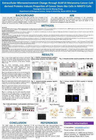
Fig. 1. These results suggest that the B16F10 cell-derived Fig. 1. B16F10 melanoma-derived proteins
proteins with a proper concentration quickly induce colony induce colony formation from NIH3T3 cells Fig. 2. Induced-colonies acquire properties of stem cells
formation, which is a specific characteristic of stem cells,
and do not affect survival in normal mouse fibroblast
NIH3T3 cells.
Fig. 2. These experiments provide substantial
experimental evidence to support the concept that proteins
from cancer cells could construct a tumor
microenvironment that induces dedifferentiation and re-
differentiation capacities in normal cells.
Fig. 3. Our data demonstrated that the colonies induced by
the B16F10 cell-derived protein treatment in the NIH3T3
cells obtained resistance against cisplatin and mitomycin C.
This observation indicates that induced colonies acquire
the properties of CSCs such as anti-cancer drug
resistance.
Fig. 4. This observation indicates that the induced colonies Fig. 3. Induced-colonies possess specific Fig. 4. Various markers of CSCs express in induced-
acquire the CSC attributes through the induction of specific characteristics of CSCs colonies
markers, such as Oct4, Sox2, ABCG2, and CD133.
Fig. 5. This observation indicates that the induced colonies
from the NIH3T3 cells maintains the tumorigenic properties
such as solid tumor initiation, growth, and progression in
vivo, retaining their differentiating capacities.
Fig. 6. In summary, we first found that tumor-derived
protein is one of the major factors in CSCs generation,
possibly by inducing dedifferentiation of neighboring cells
surrounding the tumors. Moreover, we were able to
construct in vitro model of CSCs, by which not only
acquiring enough number of CSCs, but also the long term
culture of the cells is possible.
Fig. 5. Induced-colonies could generate solid tumor in vivo Fig. 6. Summary
CONCLUSION REFERENCES Contact information
This study argues the tremendous importance of the [1] K. Polyak, WC. Hahn, Roots and Stems: stem
extracellular microenvironmental effect on the generation of cells in cancer, Nat Med. 12 (2006) 296-300. E-mail address : gwangdoli@naver.com
CSCs. we first found that tumor-derived protein is one of the [2] C. Peitzsch, A. Tyutyunnykova, K. Pantel, A.
major factors in CSCs generation, possibly by inducing Dubrovska, The root of tumor recurrence and Phone number : 051) 200-7273
dedifferentiation of neighboring cells surrounding the tumors. metastases, Semin Cancer Biol. 44 (2017) 10-24.
Moreover, we were able to construct in vitro model of CSCs, by [3] A. Albini, M.B. Sporn, The tumour Lab of Cellomics, S11-0425, Natural Science,
which not only acquiring enough number of CSCs, but also the Dong-A university, 37, Nakdonga-Daero 550
long term culture of the cells is possible. It provides a simple microenvironment as a target for
experimental method for deriving CSCs that could be based on chemoprevention, Nat. Rev. Cancer 7 (2007) beon-gil saha-gu, Busan 49315, Korea
the development of targeted therapy techniques. 139-147.