
Page 135 - D. Cancer biology
P. 135
Study of Apoptosis in Human Lung Cancer Cells by Plasma Activation Medium
Ara Jo , Hea Min Joh , Tae Hun Chung , Jin Woong Chung 1,*
1
2
2
1 Department of Biological Sciences, Dong-A University, Busan 49315, Korea
2 Department of Materials Physics, Dong-A University, Busan 49315 Korea
BACKGROUND AIM
Non-thermal atmospheric pressure plasma (NTAPP) has been reported to have strong anti-cancer effects. We have shown that PAM produced by ME-APPJ had a significant cytotoxic effect on
Plasma is treated directly with cells or in medium or buffer for generating reactive oxygen and nitrogen A549 cells but had little effect on normal cell viability. Especially, hydrogen peroxide has
species (RONS). In this study, two kinds of coaxial transmission line resonator (CTLR) with the resonance been shown to play an important role in inducing anti-cancer effects. In this study, we
frequencies 788 MHz and 2.45 GHz was employed for plasma-medium interactions. Plasma treatment present the rationale for the fundamental effect of PAM on cancer cells. Cell death of
generated the reactive species in gas phase, and these species dissolve into the liquid phase, resulting in A549 cells was mainly via apoptosis, which occurred due to a higher level of intracellular
the appearance of RONS in liquid. The concentration of H2O2 and NO2- in the plasma-activated medium ROS and was accompanied by cell cycle arrest and caspase-3/7 activation. Altogether,
(PAM) were measured by spectrophotometer. Moreover, the concentration of NO2- and H2O2 in PAM
incubating A549 cells were monitored until 24 hours. Furthermore, we measured the cell viability using these results suggest that CAP therapies based on ME-APPJ-produced PAM may be a
MTT assay. As a result, the reduced cell viability rate was observed on human lung cancer cells incubated good consideration for human lung cancer treatment.
in the PAM for 24 hours. These results suggest that two type plasma jet can generate RONS in medium
and induce the cancer cell death.
METHODS
1. Cell culture. For in vitro studies, human non-small lung cancer A549 5. Cell viability assay. In order to confirm the cytotoxicity of PAM in A549
cells. cells, we used 3-(4, 5-dimethylthiazol-2-yl)-2,5-diphenyltetrazolium bromide
(MTT; Duchefa Biochemie, Haarlem, Netherlands).
2. Plasma sources and preparation of PAM. In producing PAM, a 2.45
GHz commercial microwave-excited APPJ (PM-10, Heuermann HF- 6. Intracellular ROS detection. The percentage of intracellular ROS was
measured using Muse Oxidative Stress Assay Kit (Merck Milipore, Billerica,
Technik GmbH) was used to treat the cell culture medium. Mam USA), following the manufacturer’s protocol.
3. Griess Assay. Nitrite concentration in PAM and in PAM including 7. Annexin V & Dead staining, cell cycle and caspase 3/7 activation. To
A549 cells was determined with Griess Reagent Kit (Molecular Probes) detection apoptosis induced by PAM on A549 cells, we used MUSE
according to the manufacturer’s directions. Annexin V and Dead Cell Assay Kit.
4. Measurement of H 2 O 2 production. To measure H 2 O 2 quantification 8. Western bolt. To confirm the change in expression of genes associated
we purchased Amplex™ Red Hydrogen Peroxide/Peroxidase Assay Kit with apoptosis, we used JNK, phospho-JNK, p53, phospho-p53, Bax and
(Invitrogen, Carlsbad, USA). GAPDH.
Beate Haertel et al., Biomol. Ther. 2014 22(6), 477-490
RESULTS
Figure 1. Plasma source and plasma properties.
The ME-APPJ used for the plasma treatment on cell culture medium was
represented in Fig 1a. To confirm produced radicals by plasma, a typical
optical emission spectrum was measured from plasma and represented in Fig
1b. In Fig 1d, ME-APPJ produces the NOγ bands (200 – 300 nm), the OH
band (308 nm), the O line (777 nm), N2 emission bands (300 – 440 nm) as
well as excited Ar lines (500 – 1000 nm).
Figure 2. Measurement of nitrite concentration in PAM and PAM
incubated with A549 cells.
It was observed that NO 2 levels in the PAM with and without cells remained
–
not much changed during storage or incubation period.
Figure 3. H 2 O 2 measurement of plasma activated medium.
These results suggest that there exists a strong correlation between cell
viability and the H 2 O 2 in the PAM.
Figure 4. Cytotoxic effects of PAM on various cancer cells and normal
cells.
In figures 4, cell viability was decreased with increasing PAM incubation time.
When the cell was treated by PAM for 24 hours, the cell viability decreased
drastically but its dependence on input power and flow rate was not significant.
Figure 5. Intracellular ROS measurement of A549 cells treated with PAM.
When PAM was applied to A549 cells for 2 hours, intracellular ROS was
increased in an exposure time dependent manner. As a result, PAM can raise
intracellular ROS levels in A549 cells from 2 hours and ROS are maintained
for up to 24 hours.
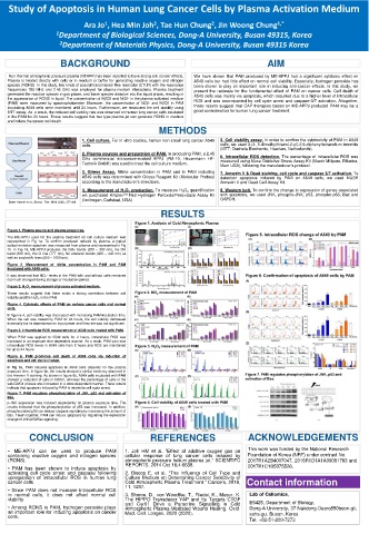
Figure 6. PAM promotes cell death of A549 cells via induction of
apoptosis and cell cycle change.
In Fig 6a, PAM induced apoptosis in A549 cells depends on the plasma
exposure time. In figure 6b, the results showed a similar tendency observed in
the Annexin V staining. As shown in figure 6c, A549 cells incubated with PAM
showed a reduction of cells in G0/G1, whereas the percentage of cells in the
sub-G0/G1 phases was increased in a dose-dependent manner. These results
indicate that apoptosis induced by PAM is related to cell cycle arrest.
Figure 7. PAM regulates phosphorylation of JNK, p53 and activation of
Bax.
p-JNK expression was induced dependently on plasma exposure time. The
results indicated that the phosphorylation of p53 was increased. In addition,
phosphorylated p53 can induce caspase signaling by increasing the amount of
Bax. Taken together, PAM can induce apoptosis by regulating the expression
change of JNK/p53/Bax signaling.
CONCLUSION REFERENCES ACKNOWLEDGEMENTS
• ME-APPJ can be used to produce PAM 1. Joh HM et al. ”Effect of additive oxygen gas on This work was funded by the National Research
containing reactive oxygen and nitrogen species cellular response of lung cancer cells induced by Foundation of Korea (NRF) under contract No.
(RONS). atmospheric pressure helium plasma jet.” SCIENTIFC 2017R1A2B4007047, 2018R1D1A1A09081763 and
• PAM has been shown to induce apoptosis by REPORTS. 2014 Oct 16;4:6638. 2017R1C1B5075526.
activating cell cycle arrest and caspase following 2. Biscop E. et al. “The Influence of Cell Type and
upregulation of intracellular ROS in human lung Culture Medium on Determining Cancer Selectivity of
cancer cells. Cold Atmospheric Plasma Treatment." Cancers, 2019, Contact information
• Since PAM does not increase intracellular ROS 11, 1287.
in normal cells, it does not affect normal cell 3. Shome, D., von Woedtke, T., Riedel, K., Masur, K. Lab of Cellomics,
viability. The HIPPO Transducer YAP and Its Targets CTGF E5425, Department of Biology,
• Among RONS in PAM, hydrogen peroxide plays and Cyr61 Drive a Paracrine Signalling in Cold Dong-A University, 37 Nakdong-Dearo550beon-gil,
Atmospheric Plasma-Mediated Wound Healing. Oxid.
an important role for inducing apoptosis on cancer Med. Cell. Longev. 2020 (2020). saha-gu, Busan, Korea
cells.
Tel: +82-51-200-7273