
Page 15 - C. Biotechnology and molecular imaging
P. 15
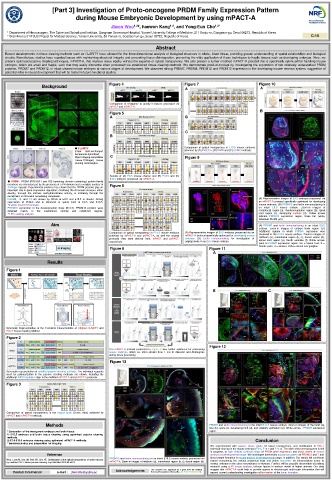
[Part 3] Investigation of Proto-oncogene PRDM Family Expression Pattern
during Mouse Embryonic Development by using mPACT-A
Jiwon Woo 1,2# , haewon Kang , and Yong-Eun Cho 1,2*
1,2
1 Department of Neurosurgery, The Spine and Spinal cord Institute, Gangnam Severance Hospital, Yonsei University College of Medicine, 211 Eonju-ro, Gangnam-gu, Seoul 06273, Republic of Korea
2 Brain Korea 21 PLUS Project for Medical Science, Yonsei University, 50 Yonsei-ro, Seodaemun-gu, Seoul 03722, Republic of Korea C-10
Abstract
Recent developments in tissue clearing methods such as CLARITY have allowed for the three-dimensional analysis of biological structures in whole, intact tissue, providing greater understanding of spatial relationships and biological
circuits. Nonetheless, studies have reported issues with maintaining structural integrity and preventing tissue disintegration, preventing the wide application of these techniques to fragile tissues such as developing embryos. Here, we
present optimized passive clearing techniques, mPACT-A, that improve tissue rigidity without the expense of optical transparency. We also present a further modified mPACT-A protocol that is specifically optimized for handling mouse
embryos, which are small and fragile, such that they easily dismantle when processed via established tissue clearing methods. We demonstrate proof-of-concept by investigating the expression of two relatively understudied PRDM
proteins, PRDM7 and PRDM12, in intact cleared mouse embryos at various stages of development. We observed strong PRDM7, PRDM8, PRDM12 and PRDM13 expression in the developing mouse nervous system, suggestive of
potential roles in neural development that will be tested in future functional studies.
Figure 4 Figure 7 Figure 10
Background
A A
Comparison of resistance to gravity in tissues processed via
mPACT and mPACT-A.
B
Figure 5
A B
C
B
● CLARITY Comparison of optical transparency in E13.5 mouse embryos
(Clear Lipid-exchanged achieved by (A) iDISCO+, (B) BABB and (C) CUBIC methods.
Acrylamide-hybridized
Rigid Imaging-compatible C Figure 9
Tissue-hYdrogel) tissue
clearing technologies.
Results of (A) E8.5 mouse chorion and (B) E10.5 and (C)
E15.5 embryos processed via mPACT-A.
● PRDM : PRDM (PRDI-BF1 and RIZ homology domain containing) protein family
members are characterized by the presence of a PR domain and a variable number of Figure 6
Zn-finger repeats. Experimental evidence has shown that the PRDM proteins play an
important role in gene expression regulation, modifying the chromatin structure either
directly, through the intrinsic methyltransferase activity, or indirectly through the
recruitment of chromatin remodeling complexes.
PRDM8, 12, and 13 are shown by WISH at E9.5 and E10.5 in mouse. Strong (A) Representative images of E9.5 embryos processed via
expression of Prdm8 and is observed in spinal cord at E9.5 and E10.5. an mPACT-A protocol specifically optimized for developing
whole mount ISH analysis. mouse embryos. (B) PRDM12 and lectin immunostaining in
PRDM10 expression in the developmental stage (E13.5). PRDM10 proteins were an intact E9.5 mouse embryo. Zoom-in images of
detected mainly in the craniofacial, somital and notochord regions. craniofacial region (a), rhombencephalon region (b), spinal
IHC staining analysis. cord region (c), developing somites (d). Yellow arrows
indicate PRDM12 expression region. Scale bar (white,
individual 50-300 μm).
Aim (A) PRDM7 and lectin immunostaining in an intact E9.5
embryo. Zoom-in images of embryo body region. (B)
Comparison of optical transparency in E13.5 mouse embryos (A) Representative images of E8.5 embryos processed via an Additional regions in which PRDM7 expression was
observed in the E10.5 mouse embryo. Zoom-in images of
achieved by mPACT-A and psPACT-A, as well the original mPACT-A protocol specifically optimized for developing mouse midbrain (a), craniofacial region (b, c), dorsal region (d),
protocols they were derived from, mPACT and psPACT, embryos. (B) Lectin immunostaining for investigation of spinal cord region (e), fourth ventricle (f). White arrows
respectively. angiogenesis in an E8.5 mouse embryo.
point to PRDM7 expression region. SC = Spinal cord; S =
Somite pairs; A = amnion; DRG = dorsal root ganglion.
Figure 8 Figure 11
A
Results
Figure 1
B C
Schematic Representation of the Technical Characteristics of Original CLARITY and
PACT Tissue Clearing Method.
Figure 2
Figure 12
The mPACT-A method presented in Figure 2 was further optimized for processing
mouse embryos, which are often smaller than 1 mm in diameter and disintegrate
during tissue processing.
Figure 13
Schematic representation of modified passive clearing methods. The individual reagents
used for polymerization in the passive clearing methods are shown, including the
additional A4P0 incubation step in the modified mPACT-A and psPACT-A protocols.
Figure 3
Comparison of optical transparency in the mouse brain (2-mm thick) achieved by
mPACT and mPACT-A methods.
Methods PRDM7 and lectin immunostaining in the intact E13.5 mouse embryo. Zoom-in images of the heart (a),
liver (b), spine (c), developing limb (d), and olfactory epithelium (e). White arrows : PRDM7 expression
* Generation of the transparent embryos and brain tissue region.
1) E13.5 embryos and brain tissue clearing using optimized passive clearing
methods.
2) E7.5-E11.5 embryos clearing using optimized mPACT-A methods. Conclusion
* Immunostaining and preparation for imaging
We experimented with passive tissue clarity for tissue transparency, and modification of PACT
technical clearing in the mouse embryos (E9.5, E10.5 and E13.5). The work of tissue transparent is still
in progress, to high visibility confocal image of PRDM gene expression and blood vessel of mouse
Reference embryos in developmental stage. We investigate extensively expression pattern of PRDM12 and 7 and
PRDM12 and lectin immunostaining in an intact E10.5 mouse embryo processed via blood vessel formation in mouse embryo of developmental stage. In addition, Our results will contribute
Woo J, Lee M, Seo JM, Park HS, Cho YE. Optimization of the optical transparency of rodent tissues toward overcoming the various preclinical trials and define biomarker for intramedullary tumor to
by modified PACT-based passive clearing. Exp Mol Med 2016;48:e274. mPACT-A. Zoom-in images of midbrain (a), craniofacial region (b, c), dorsal region (d).
various embryos in the close resemblance to humans. Further, will be possible observation of probing
research using a 3D image analysis, inducer factors in embryo model of higher animals. Our data
Acknowledgements This research was supported by a grant from the National suggest that mPACT-A could help to provide access to stereoscopic multi-scale information that will
Contact Information e-mail : jiwonflu@yuhs.ac Research Foundation of Korea (No. 2017R1D1A1B03030315). expand current understanding investigation of bio-marker of the tumor formation.