
Page 19 - C. Biotechnology and molecular imaging
P. 19
Dynamic Visualization of Osmotic Stress-Induced Redox Reaction in
Nucleus by FRET-based Redox biosensor
Jung-Soo Suh1, Yoon-Kwan Jang1, Heon-Su Kim1, Sang-Hyun Ahn1 and Tae-Jin Kim1, 2*
1 Department of Integrated Biological Science, Pusan National Unversity, Pusan 46241, Korea (Republic of)
2 Department of Biological Sciences, Pusan National Unversity, Pusan 46241, Korea (Republic of)
Abstract Purpose
Redox reactions are critical for numerous cellular processes, including energy metabolism, To investigate the molecular mechanism how redox activities can
cell cycle, gene expression and regulation. However, it remains unclear how redox activity be regulated in subcellular organelles during osmotic stress
is regulated in the subcellular organelle. In this study, we have developed a nucleus- • Osmotic stress-induced cellular damage remains insufficient for
targeted redox biosensor based on fluorescence resonance energy transfer to visualize pathological cell signaling.
the spatiotemporal dynamics of redox reactions in the nucleus. Our results reveal that salt- • Mechanochemical factors such as osmotic stress have an
induced osmotic stress stimulates nuclear redox reactions, but a similar event is rarely influence on cellular processes which are essential for cell cycle,
detected in the nucleus of H2O2-treated cells. We further demonstrate that redox stress gene expression and regulation.
tolerance occurs only in the nucleolus, known as the main site of ribosome biogenesis, as • However, little is known how redox event is regulated in the
compared to the nucleoplasm region, which could be due to aggregation of peroxiredoxin- nucleus and nucleolus due to limitations in experimental
I. Thus, our data suggest that salt-induced redox stress acts to the minimum in the approaches.
nucleolus, membrane-less organelles where antioxidants can serve as alternative To visualize the dynamics of salt-induced redox stress with high
protection against osmotic/redox stress. spatiotemporal resolution through FRET-based live-cell imaging
RESULTS
Figure 1 Figure 2 Figure 3
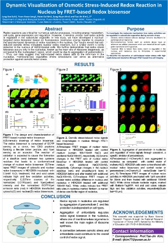
N orm a liz e d C FP /FR E T R a tio 1 .6 1 .4 1 .2 1 .0 C o n oidinA (1 µM ) pretrea ted N ucle olar R egion
-1 0 0 10 20 30 N on-N ucleolar R egion
Time(min)
CONCLUSION
ACKNOWLEDGEMENTS
This research was supported by Basic Science
Research Program through the National Research
Foundation of Korea (NRF) funded by the Ministry
of Education (2017R1D1A1B03035622).
Contact information
* Correspondence: Prof.Tae-Jin Kim,
E-mail: tjkim77@pusan.ac.kr