
Page 11 - C. Biotechnology and molecular imaging
P. 11
[Part 1] Comparison of Clearing Efficacies of Various Tissue Clearance Protocols
in Rodent Brain
Jiwon Woo 1,2# , Mirae Lee , Jeong-Yoon Park , and Yong-Eun Cho 1,2*
1,2
1,2
1 Department of Neurosurgery, The Spine and Spinal cord Institute, Gangnam Severance Hospital, Yonsei University College of Medicine, 211 Eonju-ro, Gangnam-gu, Seoul 06273, Republic of Korea
2 Brain Korea 21 PLUS Project for Medical Science, Yonsei University, 50 Yonsei-ro, Seodaemun-gu, Seoul 03722, Republic of Korea C-8
Abstract
The mammalian biological systems such as the central nervous system including brain and spinal cord consist of over thousands of distinct cell types forming widely interconnected functional neural networks, and coordinated across
multiple size scales, from the sub-nanoscale of molecules to the macroscale, the tissue wide interconnectivity of the cell populations. Here we introduce comparative analyses of clearing efficacies in various tissue clearing techniques for
high resolution imaging analysis of the multiscale organization of intact brain tissues in rodent. We evaluated clearing efficacies of the brain tissue using improved methods with twenty passive clarity methods that allowed production of
transparent brain by tissue clearing. The tissue comparative analysis facilitates the rapid examination of three dimension morphological and therapeutic aspects of surgical animal disease models and can be selective used to aid in the
investigating of medical and experimental conditions. Our comparative analyses of tissue clearing efficacies suggest that useful to provide access to adaptable method for stereoscopic multi-scale information that will expand current
understanding of health and disease.
Background Figure 4 Figure 7
● CLARITY
(Clear Lipid-exchanged Acrylamide-hybridized Rigid Imaging-compatible Tissue-
hYdrogel) tissue clearing technologies.
Results
Figure 1
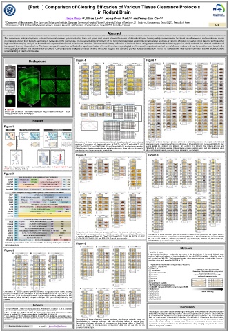
Comparison of tissue clearance process achieved via gelation-based tissue clearing Comparison of tissue clearance process achieved in rat brains processed via organic solvent-based
protocols. Comparison of clearing efficacies of PACTs (psPACT and mPACT) (A), clearing protocols. Comparison of clearing efficacies of Ethanol BABB (A), 1-propanol BABB (B), tert-
SWITCHs (SWITCH-1 and SWITCH-4) (B), and Tissue-MAP (C) in rodent brain samples. butanol BABB (C), 3DISCO (D), iDISCO+ (E), uDISCO (F), FDISCO (G), Ethanol-ECi (H), and
Optical images showing samples before and after clearance, along with any changes in PEGASOS (I) on rat brain samples. Optical images showing samples before and after clearance, along
sample size upon tissue processing, are included. with any changes in sample size upon tissue processing, are included.
Figure 5 Figure 8
Schematic Representation of the Technical Characteristics of Original CLARITY and
PACT Tissue Clearing Method.
Figure 2
Comparison of tissue clearance process achieved via clearing methods based on
hyperhydration or aqueous solutions with high-refractive indices in rat brain. Comparison Comparison of tissue clearance process achieved in mouse brains processed via organic solvent-
of clearing efficacies of Clear T (A), Clear T2 (B), ScaleA2 (C), CUBIC (D), FOCM (E), RTF based clearing protocols. Comparison of clearing efficacies of Ethanol BABB (A), 1-propanol BABB
(F), SeeDB (G), SeeDBp (H), and 80% TDE (I) on rat brain samples. (B), tert-butanol BABB (C), 3DISCO (D), iDISCO+ (E), uDISCO (F), FDISCO (G), Ethanol-ECi (H),
and PEGASOS (I) on mouse brain samples.
Schematic representation of the 24 protocols of the 17 clearing techniques used in this Figure 6
comparative study.
Methods
Figure 3
* Isolation of tissue
Upon opening the thorax, an incision was made to the right atrium of the heart. Animals were
perfused with equal volumes of heparin dissolved in ice-cold PBS (10 units/mL; 50 mL for rats, 20
mL for mice) and 4% PFA. The brain was isolated using general methods, cut into either 2 mm or 4
mm-thick slices, and placed in 4% PFA at 4°C.
* Preparation of rodent brain samples tissue clearance
(1) psPACT and mPACT
(2) SWITCH
(3) Tissue-MAP
(4) ClearT and ClearT2
(5) ScaleA2
(6) CUBIC
(7) FOCM
(8) RTF
(9) SeeDB and SeeDBp
(10) Thiodiethanol-based clearing
(11) BABB, 1-propanol BABB and tert-butanol BABB
(12) 3DISCO
Comparison of tissue clearance process achieved via gelation-based tissue clearing (13) iDISCO+
protocols in mouse brain. Comparison of clearing efficacies of psPACT (A), mPACT (B), (14) uDISCO
and SWITCH-1 (C) in mouse brain samples. Optical images showing samples before and (15) FDISCO
after clearance, along with any changes in sample size upon tissue processing, are (16) Ethanol-ECi
included. (17) PEGASOS
Reference
Conclusion
* Chung K, Wallace J, Kim SY, Kalyanasundaram S, Andalman AS, Davidson TJ, et al. Structural
and molecular interrogation of intact biological systems. Nature 2013;497:332-7. This suggests that future studies attempting to investigate three-dimensional cerebellar structure
* Woo J, Lee M, Seo JM, Park HS, Cho YE. Optimization of the optical transparency of rodent using tissue clearing methods might benefit from further optimization of CLARITY-based protocols
tissues by modified PACT-based passive clearing. Exp Mol Med 2016;48:e274.
* Ku T, Swaney J, Park JY, Albanese A, Murray E, Cho JH, et al. Multiplexed and scalable super- as to maximize the resolution of structural information that can be obtained. This in turn fuels the
resolution imaging of three-dimensional protein localization in size-adjustable tissues. Nat Biotechnol need for future studies aimed towards improving the clearance of tissues that are hard to parse,
2016;34:973-81. Comparison of tissue clearance process achieved via clearing methods based on which arguably hold greater amounts of biological information due to increased cell and/or
hyperhydration or aqueous solutions with high-refractive indices in mouse brain. extracellular matrix density. In addition, it is provide a possible that standard transmittance
Comparison of clearing efficacies of transparent rat brain tissue tablets of Clear T (A), experiment of transparent tissue for three-dimensional deep imaging analysis at the various
Contact Information e-mail : jiwonflu@yuhs.ac ScaleA2 (B), CUBIC (C), FOCM (D), RTF (E), SeeDB (F), 60% TDE (G), and 80% TDE (H) optimum transparent conditions.
on mouse brain samples.