
Page 13 - C. Biotechnology and molecular imaging
P. 13
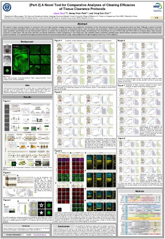
[Part 2] A Novel Tool for Comparative Analyses of Clearing Efficacies
                                        of Tissue Clearance Protocols
                                                          1,2
                                    Jiwon Woo 1,2# , Jeong-Yoon Park , and Yong-Eun Cho 1,2*


         1  Department of Neurosurgery, The Spine and Spinal cord Institute, Gangnam Severance Hospital, Yonsei University College of Medicine, 211 Eonju-ro, Gangnam-gu, Seoul 06273, Republic of Korea
     2  Brain Korea 21 PLUS Project for Medical Science, Yonsei University, 50 Yonsei-ro, Seodaemun-gu, Seoul 03722, Republic of Korea   C-9
                                                    Abstract
   The advent of tissue clearing methods, in conjunction with novel high-resolution imaging techniques, has enabled the visualization of three-dimensional structures with unprecedented depth and detail. Although a variety of clearing
   protocols have been developed, little has been done to quantify their efficacies in a systematic, reproducible fashion. Here, we present two novel assays, PACA (Punching-Assisted Clarity Analysis)-Light and PACA-Glow, which use
   easily  accessible  spectroscopy  and  gel  documentation  systems  to  quantify the transparency  of multiple  cleared tissues  simultaneously. We  demonstrate the  use  of  PACA-Light  and  PACA-Glow  to  compare  twenty tissue  clearing
   protocols on rodent brains. We also show that there are regional differences in tissue transparency in the rodent brain, with cerebellar tissue consistently achieving lower clearance levels compared to the prefrontal or cerebral cortex
   across all protocols. This represents the largest comparative study of tissue clearing protocols to date, made possible by the high-throughput nature of our PACA platforms.
                                     Figure 4                                 Figure 6
               Background                   Comparison  of tissue clearance achieved via gelation-based tissue clearing protocols.


   ● CLARITY
   (Clear  Lipid-exchanged  Acrylamide-hybridized  Rigid  Imaging-compatible  Tissue-  Comparison of clearing efficacies of Clear T  (A), ScaleA2 (B), CUBIC (C), FOCM
   hYdrogel) tissue clearing technologies.
                                                                              (D),  RTF  (E),  SeeDB  (F),  60%  TDE  (G),  and  80%  TDE  (H)  on  mouse  brain
                                                                              samples via PACA-Light.
                  Aim                Comparison of clearing efficacies of psPACT (A and B), mPACT (C and D), SWITCH-1 (E and F), SWITCH-4 (G),  Figure 7  Comparison  of  tissue  clearance  achieved  in  mouse  brains
                                                                                   processed  via organic solvent-based clearing protocols.
   Understanding the clearance properties of intact organs in a region-specific manner   and Tissue-MAP (h) in rodent brain samples (rat: 3-mm thickness, mouse: 1.5-mm thickness) via both PACA-
   can  allow  for  further  optimization  of  tissue  clearing  protocols  better  suited  to  a   Light and PACA-Glow.
   particular  region  of  interest,  and  in  turn,  a  deeper  understanding  of  the  three-
   dimensional  organization of that region that may not otherwise be uncovered.    Figure 5
                Results
   Figure 1


                                                                                            Comparison  of  clearing  efficacies  of
                                                                                            Ethanol  BABB  (A),  1P  BABB  (B),  tB
                                                                                            BABB  (C),  3DISCO  (D),  iDISCO+  (E),
                                                                                            uDISCO (F),  FDISCO (G), Ethanol-ECi
                                                                                            (H), and PEGASOS (I) on mouse brain
                                                                                            samples  (1.5-mm  thickness)  via  both
                                                                                            PACA-Light and PACA-Glow.
                                                         Comparison  of  tissue  clearance  achieved  via  clearing   Figure 8
                                                         methods based on hyperhydration or aqueous solutions
                                                         with  high-refractive  indices.  Comparison  of  clearing
                                                         efficacies of Clear T  (A), Clear T2  (B), ScaleA2 (C), CUBIC
                                                         (D), FOCM (E), RTF (F), SeeDB (G), SeeDBp (H), and
                                                         80% TDE (I) on rat brain samples (3-mm thickness) via
                                                         both PACA-Light and PACA-Glow.
                                     Figure 9
                                     A         B         E
   Schematization  of  the  Punch-based  Assistance  Clarity-measurement  Assay  (PACA)
   platform.
                                                         F
   Figure 2
   A        B                                                                                Comparison   of   tissue   clearance
                                                                                             achieved  in  rat  brains  processed  via
                        (A  and  B)  Absorbance  and                                         organic   solvent-based   clearing
                        transmittance   (%)   of   2%                                        protocols.    Comparison  of  clearing
                        agarose   gels   of   varying                                        efficacies  of  Ethanol  BABB  (A),  1-
                        thickness  (1-5mm)  at  350-850                                      propanol  BABB (B), tert-butanol BABB
                        nm.  Each  color  line  and  bar   C   D              (C), 3DISCO (D), iDISCO+ (E), uDISCO (F), FDISCO (G), Ethanol-ECi (H), and
                        point  to  assessment  values  of                     PEGASOS (I) on rat brain samples (3-mm thickness) via both PACA-Light and
                        dH2O  (DW,  blue)  and  2%                            PACA-Glow.
   C                    agarose  gel tablets  of  differing
                        thickness.  (C)  Comparison  of   G
                        light  emitted  by  the  luminous                                 Methods
                        disk.
   Figure 3        C                                    H
            B
   A
                                     Investigation of regional differences in tissue transparency achieved in rodent brains via immunofluorescence
                                     studies. Tablets were generated from three distinct regions (B1, B2, B3) of rat and mouse brain samples were
                                     processed via psPACT (A and C) or SWITCH-4 (B) or CUBIC (D). (E and F) The 3D and volumetric imaging of
   (A and B) Quantification of light transmitted from the luminous disk compared to a blank   psPACT and SWITCH-4 rat brain samples was created from serial single z-images of the blood vessel pattern
   control.  (C) Comparison  of light transmission  between  an  uncleared  4mm-thick  mouse   using a confocal microscopy in three distinct regions (B1, B2 and B3). (G and H) The 3D and volumetric imaging
   brain  and  a  sample  cleared  via  mPACT  on  a  luminous  disk  in  both  UV  and  dark   of psPACT and SWITCH-1 mouse brain samples was created from serial single z-images of the blood vessel
   conditions.                       pattern  using a confocal  microscopy  in three  distinct regions (B1, B2 and B3).
                Reference                                                                            We developed the absorbance analysis about the optically transparency
                                       Conclusion
                                                                                                     of  cleared  tissues  with  light  attenuation  quantification  using  various
   Chung K, Wallace J, Kim SY, Kalyanasundaram S, Andalman AS, Davidson TJ, et al. Structural   clearing methods of current reported. These simple PACA assays require little equipment and minimal hands on
   and molecular interrogation of intact biological systems. Nature 2013;497:332-7.
                                    analyze to transmittance  of clear  tissue  and screening the clear-agent in laboratory. In  addition, it  is provide  a
                                    possible that standard transmittance experiment of transparent tissue for three-dimensional deep imaging analysis
                                    at the various optimum transparent conditions. Our data suggest that the PACA platform including the PACA-Light
         Contact Information              e-mail :    jiwonflu@yuhs.ac   and the PACA-Glow provides access to optically information that may support to understand anatomical study.