


Page 97 - ebook
P. 97
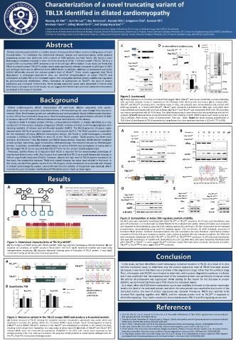
Then, chloroquine and MG132 were treated to determine which protein degradation pathway is related, and it was confirmed that
                      Characterization of a novel truncating variant of
                      TBL1X identified in dilated cardiomyopathy

                                                         1
                                                                   1
                      Hyeong-Jin Kim , Sun-Ho Lee , Sae-Bom Jeon , Hyoeun Kim , Jungyoon Choi , Jaewon Oh ,
                                                                           3
                               1,*
                                       2,*
                                                 1
                              1,2
                                          1,2
                      Ho-Geun Yoon , Sahng Wook Park , and Seung-Hyun Lee 1,2,†
                      1 Department of Biochemistry and Molecular Biology, Yonsei University College of Medicine, 50-1 Yonsei-ro, Seodaemun-gu, Seoul 03722, South Korea
                      2 Department of Biochemistry and Molecular Biology, Graduate School of Medical Science, Brain Korea 21 Project, Yonsei University College of Medicine, 50-1 Yonsei-ro, Seodaemun-gu, Seoul 03722, South Korea
                      3 Division of Cardiology, Severance Cardiovascular Hospital, Cardiovascular Research Institute, Yonsei University College of Medicine, 50-1 Yonsei-ro, Seodaemun-gu, Seoul 03722, South Korea
                      Abstract                          C                D In vitro cell-free translation assay  E
                                                          TBL1X WT
    Dilated cardiomyopathy (DCM) is a cardiac disease that causes heart failure and is a leading cause of heart
    transplantation. To investigate the relationship between disease and associated genes, whole genome
                                                          TBL1X TV
    sequencing analysis was performed with a cohort of DCM patients and their family. We found a novel
    homozygous nonsense mutation in exon 15 of the transducin β-like 1 X-linked protein (TBL1X). TBL1X is a
    subunit of the co-repressor SMRT and plays a role in the cell type differentiation. In our study, we found that  Flag-TBL1X WT  Ubiquitinated
                                                                                                       proteins
    TBL1X truncated variant (TBL1X TV ) protein levels were significantly reduced compared to wild-type in H9C2  Flag-TBL1X TV  F
    cell, although there was no difference in the mRNA levels. In addition, inhibition of proteasome activity by
    MG132 effectively rescued the reduced protein level of TBL1X TV . These results suggested that TBL1X
    degradation is proteasome-dependent. Also, we identified phosphorylation at serine 470/474 and
    sumoylation at Lysine 560 in the truncated region. We investigated whether protein stability was regulated
    by post-translational modification. When, restoration of sumoylation to TBL1X TV , the level of protein
    expression was increased. Furthermore, TBL1X protein expression levels were decreased in human DCM
    heart tissues compared to normal tissues. So, we suggest that TBL1X expression level and protein truncation
    are related to the cause of the DCM.
                   Background                           Figure 2. (continued)
                                                        (C) Protein expression of C-terminus-truncated FLAG-tagged TBL1X (TBL1X TV , lane 3) was confirmed in a immunoblotting
                                                        with anti-FLAG antibody. In lane 2, expression of the full-length FLAG-TBL1X protein was shown. (D) (2, 4 lanes) pSG5-
                                                        TBL1X WT and TBL1X TV constructs were translation assay in vitro, and products were immunoblotted and reacted with
     Dilated cardiomyopathy (DCM) characterized left ventricular dilation associated with systolic  FLAG-tag antibody. (1, 3 lanes) pSG5-TBL1X WT , TBL1X TV were transiently transfected into H9C2 cells, and lysates were
    dysfunction. Sometimes patients show heart failure, a life-threatening risk, even though they have been  immunoblotted and reacted with anti-FLAG antibody. (E) TBL1X TV and TBL1X WT was transfected 293FT cells. TBL1X protein
    treated. Other life-threatening risks are arrhythmias and atrioventricular blocks. Inflammatory disorders  was immunoprecipitated with M2 FLAG bead. The ubiquitinated products were detected by immunoblotting with anti-HA
    or toxic effects from alcohol or drugs cause dilated cardiomyopathy, and genetic factors affected 30-40%  antibody. (F) (Left) Representative immunohistochemistry (IHC) staining of ICMP, DCMP patient heart tissues using anti-
                                                        TBL1X antibody. Black arrows, nuclei of cardiomyocyte. Scale bar : 50μm. (Right) Bar graph showing quantification of
    of patients. About 25~35% of affected individuals have familial forms of the disease.  TBL1X level. Quantificationof TBL1X expression by ImageScope. Error bars represent the mean ± SD (n=4, **P <0.005)
     Transducin β-like 1 X-linked protein (TBL1X), a transcriptional cofactor, is initially identified as a core
    component of the nuclear receptor corepressor (NCoR) complex and also co-immunoprecipitates with
    silencing mediator of retinoic acid and thyroid receptor (SMRT). The TBL1X gene has 18 exons, spans
    approximately 150 kb of genomic sequence on chromosome Xp22.3. The NCoR complex is responsible  A  B
    for the repression of many different transcription factors. We found a novel homozygous nonsense
    mutation c.1316G>A (p.Trp439Ter) in exon 15 of the TBL1X protein. TBL1X protein has three main
    domains : LisH domain, F-box-like domain and WD40 repeat domain. Especially, WD40 domain mediated
    protein-protein interaction, signal transduction, RNA processing. This mutation site was on WD40 repeat
    domain. In addition, we identified phosphorylation at serine 470/474 and sumoylation at Lysine 560 in
    the full length TBL1X. So we expected that TBL1X TV form has the protein stability dysfunction.
     Previously, Sabrina Rivero et al reported that TBL1X is required for the mesenchymal phenotype of
    transformed breast cancer cells. Also Youssef et al reported that TBL1X as a novel therapeutic strategy in
    Diffuse large B-cell lymphoma (DLBCL). However, despite the high level of TBL1X protein expression in
    the heart, the relationship between TBL1X and related diseases has never been studied in the heart. In
    this study, we identified genetic variant in the TBL1X gene, which are likely to be associated with disease
    in Dilated cardiomyopathy patient, and confirmed that the stability of the protein was reduced, which  C
    involved the post-translation modification (PTM) site located in the truncated region.
                       Results
    A                        B                           D                      E
       Heart disease  Hepatocirrhosis
                    Hypertension
                    Diabetes mellitus
    C  DCM patient                                      Figure 3. Sumoylation at lysine 560 regulates protein stability.
                                                        (A) H9C2 cells were transiently transfected with pSG5-TBL1X WT or TBL1X TV constructs. At 24 hours post-transfection, cells
                                                        were left untreated (-) or treated with (A) 10uM MG132 for the indicated time points. Whole-cell lysates were prepared
                                                        and subjected to immunoblotting using anti-FLAG antibody against p53 and β-actin. (B) 24hr Chloroquine for the indicated
                                                        concentrations, immunoblotting using anti-FLAG antibody against LC3II and β-actin. (C) (left) Schematic structures of
                                                        truncated TBL1X proteins. Positions of phosphorylation sites and sumoylation site were indicated. (right) Serine residues
                                                        at positions 470 and 474 were mutated to alanine, Lysine residue at position 560 was mutated to Arginine. (D) H9C2 cells
                                                        were transiently transfected with pSG5-TBL1X WT or TBL1X K560R or TBL1X S470/474A constructs. At 24 hours post-transfection,
                                                        cells were left untreated (-) or treated with 10uM MG132 for 12hr. (E) (left) H9C2 cells were transiently transfected with
    Figure 1. Mutational characteristics of TBL1X p.W439*.  pSG5-TBL1X WT or TBL1X TV , or sumo-tagged TBL1X TV constructs. (right) H9C2 cells were transiently transfected with pSG5-
    (A) The Pedigree of DCMP family with TBL1X p.W439*. Black box indicated homozygous affected individual. (B) IGV  TBL1X WT or TBL1X K560R or sumo-tagged TBL1X K560R constructs.
    snapshot of c.1316G>A, p.Trp439* variant in TBL1X (ChrX: 9,677,677, hg19). Nonsense mutation was found using
    next-generation sequencing. (C) Sequence alignment showing point of truncation of TBL1X protein. It was highly
                                                                        Conclusion
    conserved crossing vertebrate, insect and yeast genomes.
    A
     TBL1X WT                                            In this study, we have identified a novel homozygous nonsense-mutation in TBL1X. As a result of in vitro
                                    TBL1X WT
                                                        cell-free translation assay to determine why the protein expression level of TBL1X truncated protein
                                                        decreased, it was found that there was a problem in the degradation stage rather than the synthesis stage.
                                                        Then, chloroquine and MG132 were treated to determine which protein degradation pathway is related,
     TBL1X TV                                           and it was confirmed that the expression level of the truncated protein was particularly increased when
                                    TBL1X TV            the activity of proteasome was suppressed. While looking for the reason for the decrease in protein
                                                        stability, we confirmed that there was a PTM sites in the truncated region.
                                                         As a result, when the K560 where sumoylation occurs was modified, it showed similar protein expression
    B Quantitative PCR
                     1 .5                               level to the level of the truncated protein, and when the sumo protein was attached to tne N-term of the
        2 .0  Pre-PTC  1 .5  Post-PTC  N o n - t r e a t  truncated protein, the level of protein expression was restored. Previously, TBL1X was reported to be
       R e la tiv e  e x p r e s s io n /G A P D H  1 .5 1 .0 0 .5  R e la tiv e  e x p r e s s io n /G A P D H R e la tiv e  e x p r e s s io n /G A P D H  1 .0 0 .5 1 .0  involved Wnt signaling together with TBLR1, and the reduced protein level by TBL1X TV is expected to
                                C H X
                                                        affect Wnt signaling. Thus, further studies for interaction between TBL1X and Wnt signaling are needed.
                     0 .5
        0 .0
         T B L 1 X -W T  T B L 1 X -T V  0 .0 0 .0 B L 1 X -W T  T B L 1 X -T V  References
                     T
                      T B L 1 -W T
                           T B L -T V
                                                         Wnt signaling.Mol Cell. 2011;43(2):203-16.
    Figure 2. Nonsense variant in the TBL1X escape NMD and produce a truncated protein.  1. Choi HK, Choi KC, Yoo JY, Song M, Ko SJ, Kim CH, et al. Reversible SUMOylation of TBL1-TBLR1 regulates beta-catenin-mediated
    (A) Domain structure of TBL1X showing the nonsense mutation introduced a premature stop codon which was  2. Tatsuaki Kurosaki, Maximilian W. Popp & Lynne E. Maquat. Quality and quantity control of gene expression by nonsense-mediated
                                                         mRNA decay. Nat Rev Mol Cell Biol. 2019;20(7):406-420.
    predicted to truncate the protein within the C-terminal WD40 repeat domain. TBL1X chromatograms of normal  3. Pohl C, Dikic I. Cellular quality control by the ubiquitin-proteasome system and autophagy. Science. 2019;366(6467):818-822.
    (TBL1X WT ) and p.W439* (TBL1X TV ). Cytosine in the TBL1X WT was substituted to Thymine in the mutant (red box),  4. John Lynn Jefferies MD, Prof Jeffrey A Towbin MD. Dilated cardiomyopathy. Lancet. 2010;375(9716):752-62
    resulting in the change from Tryptophan to a stop codon at amino acid 439. (B) Levels of TBL1X WT and TBL1X TV -PTC  5. Annie Turkieh, Sina Porouchani, Olivia Beseme, Maggy Chwastyniak, Philippe Amouyel, Nicolas Lamblin, et al. Increased clusterin
    related mRNAs were also measured by quantitative PCR(qPCR) in the H9C2 cells. Values were expressed as fold  levels after myocardial infarction is due to a defect in protein degradation systems activity. Cell Death Dis. 2019;10(8):608
    changes (means ± SD, n=3). Cells were treated in the presence of PBS (control) or cycloheximide (100ug/ml) for 2h.  6. Ya-Ting Wang, Jocelyn Chen, Chou-Wei Chang, Jayu Jen, Tzu-Yu Huang, et al. Ubiquitination of tumor suppressor PML regulates
    Pre-PTC, Post-PTC means primer binding location.     prometastatic and immunosuppressivetumor microenvironment. J Clin Invest. 2017;127(8):2982-2997

