


Page 101 - ebook
P. 101
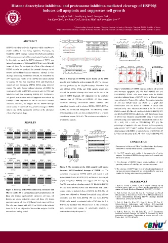
Histone deacetylase inhibitor- and proteasome inhibitor-mediated cleavage of HSP90β
                            induces cell apoptosis and suppresses cell growth

                                      Sangkyu Park , Jae-Hyung Jeon , Jeong-A Park ,
                                                                       1
                                                            2
                                                1
                                Jun-Kyu Choi , Ye-Ram Choi , Ha-Eun Shin and Younghee Lee 1, 2*
                                                    2
                                         2
                                                              2
                                                 1 Biotechnology Research Institute, Chungbuk National University, Cheongju, Chungbuk 28644, Republic of Korea
                                     2 Department of Biochemistry, College of Natural Sciences, Chungbuk National University, Cheongju, Chungbuk 28644, Republic of Korea
                                                                                                        .
                ABSTRACT
     HSP90 is one of the molecular chaperones which contributes to
     protein stability in most living organisms. Previously, we
     found that HSP90 cleavage occurred when histone deacetylase
     inhibitor or proteasome inhibitor were treated in leukemia cells.
     In this study, we found that HSP90 cleavage of HSP90 was
     induced by treatment of SAHA and MG132 in 6 out of 16 solid
     tumor cell lines. To investigate the effects of the cleavage of
     HSP90 on cells, we predicted the potential cleavage site of
     HSP90 and established mutant constructs. Through in vitro
     cleavage assay using recombinant proteins, we found that the
     294 th aspartic acid residue of the HSP90β was mainly cleaved  Figure 2. Cleavage of HSP90β occurs mainly at the 294th
     by caspase 10. We then established K562 and Mia-PaCa-2  aspartic acid residue by active caspase 10. (A) The cleavage
     mutant cell lines expressing HSP90β D294A using retroviral  site was predicted by the CaspDB database. Thereafter, four
     system. The cells showed reduced cleavage of HSP90 by  sites (252nd, 259th, 278th, and 294th aspartic acids) were  Figure 4. Inhibition of HSP90 cleavage reduces cell growth
     treatment of SAHA and MG132 compared with the K562 and  selected for potential cleavage sites based on the size of the  and increases apoptosis. (A) The K562-HSP90β WT and
     Mia-PaCa-2 cell lines expressing HSP90β WT. Furthermore,  cleaved HSP90 fragment. (B) Alignment for the predicted  K562-HSP90β D294A cells were treated with the DMSO
     cell growth was increased and cell apoptosis was reduced by  cleavage sites of HSP90α and HSP90β. (C) Four mutant  control or indicated doses of MG132 for 24 h. The cell lysates
     HSP90β D294A expression in SAHA- and MG132-treated                    were subjected to Western blot analysis. The relative intensities
     condition. Therefore, we suggest that the HSP90 cleavage  constructs  encoding  recombinant  human  HSP90β  were  of Akt and GSK3β bands are shown as a graph after
     widely occurs in several cell lines, and the cleavage of HSP90  established (aspartic acid to alanine; D252A, D259A, D278A,  normalization with the levels of GAPDH. P values were
                                                                           evaluated using t test. Values are the mean ± SD. *P < 0.05 vs
     may be one of the mechanisms involved in the anti-tumor  D294A) by site-directed mutagenesis. The rhHSP90β mutant  DMSO control. (B) The cell viability of the K562-HSP90β WT
     effects of anti-cancer drugs.      proteins produced in E. coli were incubated with 2 U of active  and K562-HSP90β D294A cells treated with the indicated doses
                                        recombinant caspase 10 for 4 h. The mixtures were subjected to  of MG132 was measured using the MTS assay. P values were
                                        Western blot analysis.             evaluated using a ratio paired t test. Values are the means ± SD.
                 RESULTS                                                   *P < 0.05, **P < 0.01 vs. K562-HSP90β WT. (C) The
                                                                           percentages of apoptotic cells were detected by flow cytometry
                                                                           using annexin V-FITC and PI staining. The cells were obtained
                                                                           after treatment with DMSO or indicated doses of MG132 for 24
                                                                           h. Values are the means ± SD. *P < 0.05 vs. K562-HSP90β WT.

                                                                                     CONCLUSION
                                                                           1. Proteasome inhibitor and HDAC inhibitor induce the cleavage
                                                                            of HSP90 in various tissue-derived cancer cell lines.
                                                                           2. The 294 th aspartic acid residue is the major site of active
                                                                            caspase 10-mediated HSP90 cleavage in the cells treated with
                                                                            MG132 or SAHA.
                                                                           3. The cleavage of HSP90 induces down-regulation of client
                                                                            proteins, reduces cell viability, and increases apoptosis.
                                        Figure 3. The mutation at the 294th aspartic acid residue
                                                                           4. The cleavage of HSP90 may be a novel action mechanism of
                                        decreases MG132-mediated cleavage of HSP90. (A) The  various anti-cancer drugs and has a potential to be used as a
                                        expression of exogenous HSP90β mRNA and protein in cells  marker for finding anti-cancer drug candidates.
                                        was determined using RT-PCR (left) and Western blot analysis
                                        (right). Exogenous HSP90β was tagged with the Myc-tag.  REFERENCES
                                        GAPDH was used as a loading control. (B) The K562-HSP90β
                                                                           1. Beck R, Verrax J, Gonze T, et al. Hsp90 cleavage by an
                                        WT and K562-HSP90β D294A cells were treated with DMSO  oxidative stress leads to its client proteins degradation and
     Figure 1. Cleavage of HSP90 is induced by treatment with  vehicle control or indicated doses of MG132 for 24 h. The cell  cancer cell death. Biochem Pharmacol. 2009;77(3):375-383.
     MG132 and SAHA in various tissue-derived solid tumor cell  lysates were subjected to Western blot analysis using indicated  2. Park S, Park JA, Jeon JH, Lee Y. Traditional and Novel
     lines. (A) Human hepato-cellular carcinoma cell lines. (B)            Mechanisms of Heat Shock Protein 90 (HSP90) Inhibition in
                                                                           Cancer Chemotherapy Including HSP90 Cleavage. Biomol
     Human and mouse colorectal cancer cell lines. (C) Human  antibodies. (C) The K562-HSP90β WT and K562-HSP90β  Ther (Seoul). 2019;27(5):423-434.
     pancreatic cancer cell lines. (D) Human breast cancer cell lines.  D294A cells treated or untreated with z-VAD-fmk for 1 h,  3. Park S, Park JA, Kim YE, Song S, Kwon HJ, Lee Y.
     The cells were treated with MG132 or SAHA at the indicated  followed by treatment with MG132 for 24 h. The cell lysates  Suberoylanilide hydroxamic acid induces ROS-mediated
                                                                           cleavage of HSP90 in leukemia cells. Cell Stress Chaperones.
     doses for 24 h and subjected to Western blot analysis. GAPDH  were incubated with caspase 10 colorimetric substrate to  2015;20(1):149-157.
     was used as a loading control.     measure the activity of caspase 10.  4. Park S, Park JA, Yoo H, Park HB, Lee Y. Proteasome
                                                                           inhibitor-induced cleavage of HSP90 is mediated by ROS
                                                                           generation and caspase 10-activation in human leukemic cells.
                                                                           Redox Biol. 2017;13:470-476.

