


Page 95 - ebook
P. 95
Digging into N-glycosylation
in Human Brain tissue and Cerebrospinal Fluid
1,2
1,2
1,2
1,2
Sol Kim , Dea Sik Cho , Heejin Jeong , Myung Jin Oh , and Hyun Joo An 1,2*
1 Asia-Pacific Glycomics Reference Site, Chungnam National University, Daejeon, Korea
2 Graduate School of Analytical Science and Technology, Chungnam National University, Daejeon, Korea
Overview Result
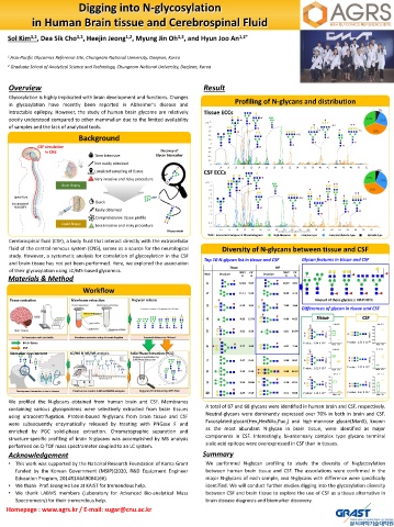
Glycosylation is highly implicated with brain development and functions. Changes
Profiling of N-glycans and distribution
in glycosylation have recently been reported in Alzheimer's disease and
intractable epilepsy. However, the study of human brain glycome are relatively Tissue ECCs
poorly understood compared to other mammalian due to the limited availability
of samples and the lack of analytical tools.
Background
CSF circulation
in CNS Discovey of
Time Intensive Glycan biomarker
Not easily obtained
Localized sampling of tissue CSF ECCs
Very invasive and risky procedure
Brain Biopsy
Spinal Cord
Quick
Cerebrospinal
fluid (CSF)
Easily obtained
Comprehensive tissue profile
Liquid Biopsy
Less invasive and risky procedure
Glycoprotein
*ECC : Extracted Compound Chromatogram ■ High Mannose ■ Complex type ■ Complex/Hybride type ■ Hybride type
Cerebrospinal fluid (CSF), a body fluid that interact directly with the extracellular
fluid of the central nervous system (CNS), serves as a source for the neurological Diversity of N-glycans between tissue and CSF
study. However, a systematic analysis for correlation of glycosylation in the CSF
Top 10 N-glycan list in tissue and CSF Glycan features in tissue and CSF
and brain tissue has not yet been performed. Here, we explored the association
Tissue CSF
of their glycosylation using LC/MS-based glycomics.
Rank Structure NAPI CV Structure NAPI CV
%
%
%
%
Materials & Method
1 12.63 6.07 10.57 3.01
Workflow 2 11.11 9.40 9.40 2.12
Tissue sonication Membrane extraction N-glycan release Amount of those glycans ≥ NAPI 40 %
3 6.50 3.64 7.90 3.66
Differences of glycan in tissue and CSF
4 4.82 13.79 4.96 0.93 Tissue CSF
7_6_4_2 5_4_0_1
X3
4 2.5
5 3.91 9.93 4.61 10.79 3 2.0
NAPI (%) 2 NAPI (%) 1.5
1.0
1
0.5
Brain tissue P-value : 1.84 X 10 -3 0 P-value : 1.32 X 10 -11 0.0
6 3.77 1.90 3.97 7.36 Tissue CSF Tissue CSF
CSF 5_4_0_2
X2 6_6_3_1
Biomarker development LC/MS & MS/MS analysis Solid Phase Extraction (PGC) 3 4
3
2
7 3.27 0.44 2.89 8.67 NAPI (%) NAPI (%) 2
1
1
P-value : 2.62 X 10 -4 0 P-value : 2.37 X 10 -2 0 Tissue CSF
8 3.13 0.93 2.88 16.51 Tissue CSF
5_6_2_1 5_4_1_2
4.5 1.0
4.0 0.8
9 2.95 10.86 2.58 0.55 3.5 0.6
NAPI (%) 3.0 NAPI (%) 0.4
2.5 0.2
10 2.86 8.59 2.51 0.50 P-value : 1.14 X 10 -5 2.0 P-value : 2.67 X 10 -2 0.0
Tissue CSF Tissue CSF
We profiled the N-glycans obtained from human brain and CSF. Membranes
A total of 87 and 68 glycans were identified in human brain and CSF, respectively.
containing various glycoproteins were selectively extracted from brain tissues
Neutral-glycans were dominantly expressed over 70% in both in brain and CSF.
using ultracentrifugation. Protein-bound N-glycans from brain tissue and CSF
Fucosylated-glycan(Hex HexNAc Fuc ) and high-mannose glycan(Man5), known
were subsequently enzymatically released by treating with PNGase F and 3 5 1
as the most abundant N-glycan in brain tissue, were identified as major
enriched by PGC solid-phase extraction. Chromatographic separation and
components in CSF. Interestingly, bi-antennary complex type glycans terminal
structure-specific profiling of brain N-glycans was accomplished by MS analysis
sialic acid epitope were overexpressed in CSF than in tissues.
performed on Q-TOF mass spectrometer coupled to an LC system.
Acknowledgement Summary
• This work was supported by the National Research Foundation of Korea Grant We performed N-glycan profiling to study the diversity of N-glycosylation
funded by the Korean Government (MSIP)(2020, R&D Equipment Engineer between human brain tissue and CSF. The associations were confirmed in the
Education Program, 2014R1A6A9064166). major N-glycans of each sample, and N-glycans with difference were specifically
• We thank Prof. Jeong Ho Lee at KAIST for tremendous help. identified. We will conduct further studies digging into the glycosylation diversity
• We thank LABMS members (Laboratory for Advanced Bio-analytical Mass between CSF and brain tissue to explore the use of CSF as a tissue alternative in
Spectrometry) for their tremendous help. brain disease diagnosis and biomarker discovery.
Homepage : www.agrs.kr / E-mail: sugar@cnu.ac.kr

