


Page 45 - ebook
P. 45
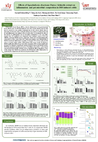
Effects of Saposhnikovia divaricata (Turcz.) Schischk extract on
inflammation and gut microbial composition in DSS-induced colitis
c
a
a
a
a,b
Saruul Erdenebileg , Yang-Ju Son , Myungsuk Kim , Da Seul Jung , Yunseong Nam ,
Nadzeya Lazerka , Chu Won Nho a,b,*
a
a Smart Farm Research Center, Gangneung Institute of Natural Products Korea Institute of Science and Technology (KIST), Gangneung, Gangwon-do 25451, Korea
b Division of Bio-Medical Science and Technology, KIST School, Korea University of Science and Technology (UST), Daejeon 34113, Korea
c Department of Food and Nutrition, Chung-Ang University, Anseong-si, Gyeonggi-do 17546, Korea
Abstract Introduction & Method
Inflammatory Bowel Disease (IBD), a chronic inflammation in the gastrointestinal
tracts, affects up to 1 in 200 individuals per year. Most current clinical drugs for IBD
lead to research for new possible treatments due to their adverse effects. Root of
Saposhnikovia divaricata (Turcz.) Schischk (SD) has been used as a medicinal herb with
its anti-inflammatory and analgesic effects. In this study, we investigated the effect of
SD extracts on inflammation and gut microbiota modulation in vitro and colitis-
induced mice. We purified seven compounds from methanol extract of SD roots Saposhnikovia
(MESD), and found MESD and two compounds; deltoin and 3-O-angeloylhamaudol, divaricata
reduced level of nitric oxide in Raw 264.7 mouse macrophages. Oral administration of Biological activities
MESD lowered pro-inflammatory cytokines, IL-1β and TNFα, in the serum and the Anti-inflammation
colon in dextran sulfate sodium (DSS)-induced IBD mouse model. Furthermore, Anti-allergic
MESD significantly shifted the gut microbial composition. DSS treatmentsignificantly
increased the abundance of pathogens and decreased the butyrate producing bacteria. Analgesic effects The FASEB Journal. 2021;35:e21439.
MESD decreased the abundance of genera Clostridium sensu stricto 1 and enriched Methodology
some of predicted metabolic activities such as chondroitin sulfate degradation. These
results suggest that MESD may ameliorate the DSS-induced colitis inflammation by • Plant extracts are rich in bioactive compounds that may act with anti-inflammatory
regulating thecompositions and functions of gut microbiota in mice. and antioxidant properties. Inhibi tion of NO production by MES D and isolated
compounds was detected by Griess reagent in Raw 264.7 cells.
• Pro-inflamma tory cytokines such as IL-1β, IL-6, and TNFα play an important role in
intestinal inflamma tion and are key factors in the trea tmen t of IBD. Inflammatory
Result cytokine levels were detected in serum from DSS -induced colitis model using a
commercial ELISA kit.
• Commensal bacterial disorders are one of the main causes of IBD. In this study, 16S
rRNA gene sequencing was used to detect the composition of the gut microbiota.
Result
A B 26
Prim-O-glucosylcimifugin 4'-O-β-D-glucosyl-5-O-methylvisamminol 5-O-Methylvisamminol Deltoin 24
B o d y w e ig h t (g ) 22 C O N
D S S
A S A
M E S D 1 0 0
20 M E S D 5 0
*
M E S D 2 0 0
18
1 2 3 4 5 6 7 8 9
5 0 # # D a y
C 4 0 D 4 0 0 # # # 4 0
S e ru m A L T (IU /L ) 2 0 ** ** T N F - (p g /m L ) 2 0 0 * IL -6 (p g /m L ) 2 0
Cimifugin 3 0 3 0 0 3 0 # # #
3-O-Angeloylhamaudol
3'-O-Acetylhamaudol
Fig 1. The structure of Prim-O-glucosylcimifugin, 4'-O-β-D-glucosyl-5-O-methylvisamminol, 5-O-Methylvisamminol, 1 0 1 0 0 1 0 * *
0
Cimifugin, Deltoin, 3'-O-Acetylhamaudol and 3-O-Angeloylhamaudol isolated from MESD. D S S 3 % - + + + + + 0 0
+
A S A (m g /k g ) - - 1 0 0 - - - D S S 3 % - + + + + D S S 3 % - + + + + +
M E S D (m g /k g ) - - - 5 0 1 0 0 2 0 0 A S A (m g /k g ) - - 1 0 0 - - A S A (m g /k g ) - - 1 0 0 - - -
-
S D sa m p le (m g /k g ) - - - S D sa m p le (m g /k g ) - - - 5 0 1 0 0 2 0 0
2 5 0 5 0 1 0 0 2 0 0
8 0 # # # 6 0 0
# #
2 0 0
P rim -O -g lu c o s y lc im ifu g in C im ifu g in 4 '-O -β -D -g lu c o s y l-5 -O -m e th y lv is a m m in o l S e ru m A S T (IU /L ) 1 5 0 6 0 4 0 *** ** *** *** 4 0 0 # # #
M E S D * IL -1 b (p g /m L ) IL -1 0 (p g /m L )
8 0 6 0 # # ## **** ** 6 0 # # ## 6 0 # # # # 60 # # ## 1 0 0 5 0 * ** *** 2 0 2 0 0
N O a m o u n t ( m o l/m L ) 4 0 2 0 **** **** N O a m o u n t ( m o l/m L ) 4 5 3 0 1 5 *** N O a m o u n t ( m o l/m L ) 4 5 3 0 1 5 ** N O a m o u n t ( m o l/m L ) 45 30 15 ** D S S 3 % 0 - - - + - - 1 0 0 + - + - 5 0 1 0 0 + - 2 0 0 + - D S S 3 % 0 - - - + - - 1 0 0 + - 5 0 + - + 1 0 0 - 2 0 0 + - D S S 3 % 0 - - - + - - + 1 0 0 - + 5 0 - 1 0 0 + - 2 0 0 + -
A S A (m g /k g )
A S A (m g /k g )
S D sa m p le (m g /k g )
S D sa m p le (m g /k g )
A S A (m g /k g )
M E S D (m g /k g )
0 0 0 0 Fig 3. MESD decreased the production of inflammatory cytokines in DSS induced mice.
C O N L P S 2 .5 5 1 0 2 0 C O N L P S 5 1 0 2 0 4 0 8 0 C O N L P S 5 1 0 2 0 4 0 8 0 C O N L P S 5 1 0 2 0 4 0 8 0 (A) Experimental design of DSS-induced colitis. (B) Changes in body weight. (C) Aspartate
aminotransferase (AST) and Alanine aminotransferase (ALT) level in serum. (D) Production of
C o n c e n t r a t io n ( g /m L ) C o n c e n t r a t io n ( M ) C o n c e n t r a t io n ( M ) C o n c e n tr a tio n ( M )
inflammatory cytokines tumor necrosis factor- α (TNF-α), interleukin-6 (IL-6), interleukin-1β (IL-1β)and
interleukin-10 (IL-10) in serum using commercial kits. Data wereexpressed as themean ± SEM (n = 7 mice
5 -O -M e th y lv is a m m in o l D e lto in 3 '-O -A c e ty lh a m a u d o l 3 -O -A n g e lo y lh a m a u d o l
per group). (*p < 0.05, **p < 0.01, ***p < 0.001, ****p < 0.0001 vs. DSS group; p < 0.05,
#
6 0 # # ## 6 0 # # ## 6 0 4 5 # # ## 6 0 4 5 # # ## ## p < 0.01 , ### p < 0.001, #### p < 0.0001 vs. control group).
N O a m o u n t ( m o l/m L ) 4 5 3 0 1 5 **** N O a m o u n t ( m o l/m L ) 4 5 3 0 1 5 **** **** **** N O a m o u n t ( m o l/m L ) 3 0 1 5 **** N O a m o u n t ( m o l/m L ) 3 0 1 5 **** **** Gut microbiota
0 0 0 0 B
C O N L P S 5 1 0 2 0 4 0 8 0 C O N L P S 5 1 0 2 0 4 0 8 0 C O N L P S 5 1 0 2 0 4 0 8 0 C O N L P S 5 1 0 2 0 4 0 8 0 A
Phylum Wilcoxon test
C o n c e n t r a t io n ( M ) C o n c e n t r a t io n ( M ) C o n c e n t r a t io n ( M ) C o n c e n tr a tio n ( M )
Fig 2. Effect of MESD and main compounds on nitricoxideproduction in vitro.
Inhibitory effect ofMESD extractand major compounds on lipopolysaccharide (LPS-1μg/mL) inducednitricoxide
(NO) production in Raw 264.7 macrophage cell. Data were expressed as the mean ±SEM (*p < 0.05, **p < 0.01,
***p < 0.001, ****p < 0.0001 vs. LPS group; # p < 0.05, ## p < 0.01 , ### p < 0.001, #### p < 0.0001 vs. controlgroup).
Conclusion
In conclusion, MESD and its isolated seven compounds decreased the
NO products in LPS induced Raw 264.7 cells. In vivo experiment, MESD Fig 4. MESD improved gut microbial composition and
reduced abundanceofClostridium sensu stricto 1
showed inhibitory effects on pro-inflammatory cytokines in serum and (A) Microbial composition at thePhylum level.
improved the symptoms of colitis and diversity of gut microbiota in DSS- (B) Differentially abundant genera between DSS and
treatmentgroups. All murine cecum samples were collected
inducedcolitis. for analyzing the microbial composition. Data were
expressed as the median ± SEM (n = 7 mice per group).
(*p < 0.05, **p <0.01, and ***p < 0.001 vs. DSS group)
This work was supported from Korea Institute of Science and Technology, Gangneung Institute of Natural Products (2Z06500)

