


Page 41 - ebook
P. 41
Monitoring of Aberrant Glycosylation in Brain
of Mouse Models of Depression using nanoLC/MS/MS
1,2
3
1,2
3
Hee Young Jo , Boyoung Lee , C. Justin Lee , Hyun Joo An *
1. Asia Glycomics Reference Site, Chungnam National University, Daejeon, 34134, Korea
2. Graduate School of Analytical Science and Technology, Chungnam National University, Daejeon, 34134, Korea
3. Center for Cognition and Sociality, Institute for Basic Science (IBS), Daejeon, 34126, Republic of Korea
Overview Result
Background Aberrant N-glycans of mouse brain in depression
The function of glycans in brain Depression specific-brain regions Backbone glycan Fucosylated and Sialylated N-glycan
Prefrontal cortex Prefrontal cortex Hippocampus
**
Social Communication
Relative Intensity (%) Relative Intensity (%)
Control Stress Control Stress
• Neurite outgrowth & fasciculation
• Synapse formation & maturation
Hippocampus
• Modulation of synaptic transmission & plasticity Higher Cognitive Process Alteration by Number of sialylation in PFC Alteration by Degree of Antennary in PFC
*N (Number of fucosylation) : 1~5
Tri-
Mono-Sial Di-Sial Mono- Di- ** Tetra-
**
** * ** *
In brain, glycans play a pivotal role in various functions depending on neural cell Relative Intensity (%) Relative Intensity (%) Relative Intensity (%)
interactions such as neurite outgrowth and fasciculation, synapse formation and
maturation, and modulation of synaptic transmission and plasticity. Recent
studies indicated that glycan is directly or indirectly related to brain disease with
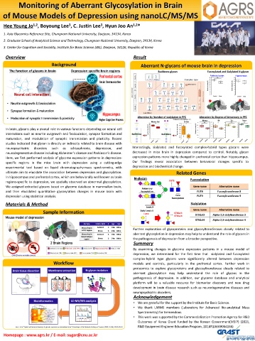
neuropsychiatric disorders such as schizophrenia, depression, and Interestingly, sialylated and fucosylated complex-hybrid types glycans were
neurodegenerative disease including Alzheimer’s disease and Parkinson’s disease. decreased in mice brain in depression compared to control. Notably, glycan
Here, we first performed analysis of glycome expression patterns in depression- expression patterns more highly changed in prefrontal cortex than hippocampus.
specific regions in the mice brain with depression using a cutting-edge Our findings reveal association between behavioral changes specific to
experimental tool based on liquid chromatography-mass spectrometry, with depression and biochemical change.
ultimate aim to elucidate the association between depression and glycosylation. Related Genes
In hippocampus and prefrontal cortex, which are behaviorally well known as brain
N-glycan Fucosylation
regions-specific to depression, we spatially observed an abnormal glycosylation. FUT8
α 6 β 4 β2 α
We assigned extracted glycans based on glycome database in mammalian brain, α 6 Gene name Alternative name
Antennary 6
and then elucidated quantitative glycosylation changes in mouse brain with fucosylation FUT8 Fucosyltransferase 8
3
depression using statistical analysis. α 3 β4 β2 α Core FUT9 Fucosyltransferase 9
3 fucosylation
α
Materials & Method Sialylation
Lewis X
Gene name Alternative name
Sialyl Lewis X
Sample Information FUT9 ST3Gal3
Mouse model of depression ST3Gal4 ST3Gal3 Alpha-2,3-sialyltransferase 3
ST3Gal4 Alpha-2,3-sialyltransferase 4
α3 α3
Sample Strain: B6/J # Age Sex
Mouse brain Control 5 14w Male Further exploration of glycoproteins and glycosyltransferases closely related to
mPFC region Stress 5 14w Male
Mouse brain Control 5 14w Male aberrant glycosylation in depression may help to understand the role of glycans in
Hippocampus
region Stress 5 14w Male the pathogenesis of depression from a broader perspective.
2 Brain Regions Summary
By examining changes in glycome expression patterns in a mouse model of
depression, we determined for the first time that sialylated and fucosylated
CVS (Chronic variable stress)
complex-hybrid type glycans were significantly altered between depression
Workflow models and controls, particularly in the prefrontal cortex. Further work in
proteomics to explore glycoproteins and glycosyltransferase closely related to
aberrant glycosylation may help understand the role of glycans in the
pathogenesis of depression. In addition, our glycome database and analytical
platform will be a valuable resource for biomarker discovery and new drug
development in brain disease research such as neurodegenerative diseases and
neuropsychiatric disorders.
Acknowledgement
• We are grateful for the support by the Institute for Basic Science.
• We thank LABMS members (Laboratory for Advanced Bio-analytical Mass
Spectrometry) for tremendous.
• This work was supported by the Commercialization Promotion Agency for R&D
Outcomes of Korea Grant funded by the Korean Government(MSIP) (2021,
Lee, J. et al. “Spatial and temporal diversity of glycome expression in mammalian brain” Proceedings of the National Academy of Sciences, 2020, 117(46), 28743-28753. R&D Equipment Engineer Education Program, 2014R1A6A9064166)
Homepage : www.agrs.kr / E-mail: sugar@cnu.ac.kr

