


Page 47 - ebook
P. 47
Senescent Tumor Cell from Colorectal Cancer
                    Accelerates Pre-metastatic Niche Formation in the Liver


                     Soon Sang Park 1,2,4* , Ga-Yeon Lee 1,4  ,Young-Kyoung Lee 1,4  ,So Hyun Park 1,3,4 , Ji Hee Park , Chan Young Lee , Dong Hyun Lee ,
                                                                           1,4
                                                                                                   1,2
                                                                                       1,2
                                                 Jang-Hee Kim 3,4†  & Tae Jun Park 1,2,4 †
                           1 Department of Biochemistry and Molecular Biology, Ajou University School of Medicine Suwon 16499, Korea;
                             2 Department of Biomedical Sciences, Ajou University Graduate School of Medicine Suwon 16499, Korea;
                                   3 Department of Pathology, Ajou University School of Medicine Suwon 16499, Korea;
                              4 Inflamm-Aging Translational Research Center, Ajou University Medical Center Suwon 16499, Korea;
                                                * Presenting Author  † Corresponding Authors
    Introduction                                               A   Figure 2                       B     Mouse IP injection model
                                                                                  Liver metastasis
                                                                 Primary colorectal cancer
                                                                                                     Control CM  Senescent CM
                                                                  p16 INK4A  p16 INK4A Tumor  CD11b  Tumor
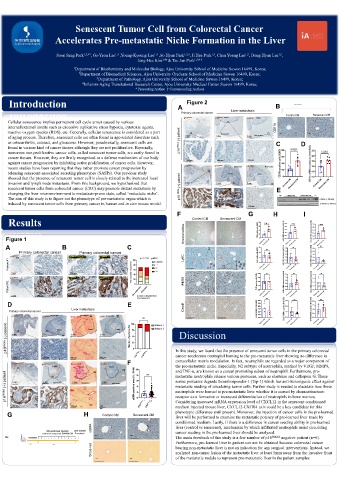
    Cellular senescence implies permanent cell cycle arrest caused by various
    internal/external insults such as excessive replicative stress hypoxia, cytotoxic agents,     CD11b
    reactive oxygen species (ROS), etc. Generally, cellular senescence is considered as a part   Non-tumor  200 μm  200 μm
    of aging process. Therefore, senescent cells are often found in age-related disorders such   5 mm  5 mm  5 mm  Non-tumor
    as osteoarthritis, cataract, and glaucoma. However, paradoxically, senescent cells are   p16 INK4A (-) patient  C  D
    found in various kind of cancer tissues although they are not proliferative. Especially,
    numerous non-proliferative cancer cells, called senescent tumor cells, are easily found in
    cancer tissues. However, they are firstly recognized as a defense mechanism of our body
                                                                  200 μm     200 μm     200 μm
    against cancer progression by inhibiting active proliferation of cancer cells. However,
                                                                  p16 INK4A  p16 INK4A  Non-tumor  CD11b  Non-tumor
    recent studies have been reporting that they rather promote cancer progression by
    releasing senescent-associated secreting phenotypes (SASPs). Our previous study   Tumor  Tumor
    showed that the presence of senescent tumor cell is closely related to the increased local
    invasion and lymph node metastasis. From this background, we hypothesized that   5 mm  5 mm  5 mm
    senescent tumor cells from colorectal cancer (CRC) may promote distant metastasis by   p16 INK4A (+) patient  E
    changing the liver microenvironment to metastasis-prone state, called ‘metastatic niche’.
    The aim of this study is to figure out the phenotype of pre-metastatic organ which is          NC  Control CM  Senescent CM  PC   CD11b (~170 kD)
    induced by senescent tumor cells from primary cancer in human and in vivo mouse model.                       α-Tubulin (~55 kD)
                                                                  200 μm     200 μm     200 μm
                                                               F                        G        H          I
                                                                    Control CM  Senescent CM
    Results
                                                                  Myeloperoxidase
   Figure 1
   A                   B                      C
        Primary colorectal cancer  Primary colorectal cancer
     p16 INK4A           p16 INK4A   Negative  p16 INK4A  1+      Ly6C
    Patient #1

     5 mm     200 μm      200 μm     200 μm
     p16 INK4A           p16 INK4A  2+  p16 INK4A  3+             F4/80
    Patient #2

     5 mm     200 μm      200 μm     200 μm      p-value is obtained from
                                                 Chi-square test
    D                                         E
                          Liver metastasis                        Clec4f
    Primary colorectal cancer
     p16 INK4A  p16 INK4A Non-tumor  Sirius Red Non-tumor  αSMA Non-tumor
   p16 INK4A (-) patient  5 mm  5 mm  Tumor  5 mm  Tumor  5 mm  Tumor  Discussion


                                                               cancer accelerates neutrophil homing to the pre-metastatic liver showing no difference in
      200 μm    200 μm    400 μm    400 μm      p16 INK4A  -  p16 INK4A +  In this study, we found that the presence of senescent tumor cells in the primary colorectal
                                              F                extracellular matrix modulation. In fact, neutrophils are regarded as a major component of
     p16 INK4A  p16 INK4A  Sirius Red  αSMA
                   Tumor     Tumor     Tumor                   the pre-metastatic niche. Especially, N2 subtype of neutrophils, marked by VEGF, MMP9,
                                                               and TNF-α, are known as a cancer promoting subset of neutrophil. Furthermore, pro-
   p16 INK4A (+) patient  5 mm  5 mm  Non-tumor  5 mm  Non-tumor  5 mm  Non-tumor  serine proteases degrade thrombospondin-1 (Tsp-1) which has anti-tumorigenic effect against
                                                               metastatic neutrophils release various proteases, such as elastease and cathepsin G. These
                                                               metastatic seeding of circulating tumor cells. Further study is needed to elucidate how these
                                                               neutrophils were homed to pre-metastatic liver whether it is caused by chemoattractant-
                                                               receptor axis formation or increased differentiation of neutrophils in bone marrow.
     200 μm     200 μm    400 μm    400                        Considering increased mRNA expression level of CXCL12 in the senescent conditioned
                                                               medium injected mouse liver, CXCL12-CXCR4 axis could be a key candidate for this
                                    μm
                                                               phenotypic difference until present. Moreover, the injection of cancer cells in the pre-learned
    G                         H      Control CM  Senescent CM
                                                               liver will be performed to examine the metastatic potency of pre-learned liver made by
                                                               conditioned medium. Lastly, if there is a difference in cancer seeding ability in pre-learned
                                 αSMA                          liver (control vs senescent), mechanism by which infiltrated neutrophils assist circulating
                 Intra-peritoneal  injection  Liver harvest    cancer seeding in the pre-learned liver should be analyzed.
                control or senescent SW480 CM  & analysis
                                  200 μm       200 μm
   Female
   Balb/c nude                                                 The main drawback of this study is a few number of p16 INK4A  negative patient (n=6).
   5wks old  Incubation
                                                               Furthermore, pre-learned liver in patient can not be obtained because colorectal cancer
                                 Sirius red                    bearing non-metastatic liver is not an indication for any surgical interventions. Instead, we
           1 week   4 weeks
                                                               analyzed non-tumor lesion of the metastatic liver at least 5mm away from the invasive front
                                                               of the metastatic nodule to represent pre-metastatic liver in the patient samples.
                                   200 μm      200 μm

