


Page 51 - ebook
P. 51
“Our findings indicate that the quality control of proteasomes
is essentially mediated by aggresomal sequestration and
subsequent autophagic degradation in mammalian cells.”
Mammalian Proteasomes Quality Control Mechanism Is Figure 1. Inhibited Proteasomes Accumulate in the Insoluble Figure 2 Inhibited Proteasomes Are Delivered to
Mediated by Ubiquitination, Aggresomal Sequestration, Fraction and the Aggresome the Aggresome through HDAC6- and Dynein-
Mediated Transport
and Autophagic Degradation
Won Hoon Choi, Seo Hyeong Park, Jiseong Kim, and Min Jae Lee*
Department of Biochemistry and Molecular Biology,
Seoul National University College of Medicine
Abstract
The 26S proteasome is a self-compartmentalized protease complex, one of
whose crucial functions is protein quality control. Multiple layers of regulatory
systems elaborately modulate proteasomal activity, i.e., hydrolysis of
polyubiquitinated proteins. However, the mechanism of destruction of
mammalian proteasomes is poorly understood. We found that inactive 26S
proteasomes are concentrated into an insoluble aggresome via HDAC6-mediated
retrograde transport. These proteasomes were colocalized with autophagic
receptor SQSTM1 in a large perinuclear inclusion body and were cleared through
selective macroautophagy, linking aggresomal segregation to proteaphagic
degradation. This pathway might be counterbalanced with recovery of
proteasomal activity and critical for reducing cellular proteasomal stress.
Structural alteration, changes in associated proteins, and polyubiquitination on
inactive proteasomes participated in the targeting mechanism to the aggresome
and autophagosome. The CHIP E3 Ub ligase polyubiquitinated purified human
proteasomes in vitro mainly with Lys63-linked polyubiquitin chains. CHIP Figure 3. Proteasomes Deposited in the Aggresome Are Cleared Figure 6. STUB1 Is Essential for Ubiquitination of
silencing significantly delayed aggresome formation by inactive proteasomes. by Autophagy Inhibited Proteasomes and Cell Survival
Thus, aggresomal sequestration and autophagic degradation are crucial for
proteasome quality control and overall mammalian protein homeostasis.
Figure 4. Proteasome Inhibitor Treatment Led to Significant
Transcriptional Up-regulation of Proteasome Subunits
Results
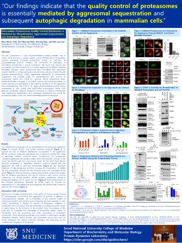
The inhibited 26S proteasome can be sequestrated in the perinuclear aggresome
(Figure 1) through HDAC6- and Dynein mediated transport (Figure 2). To
approve the fate of inhibited proteasomes accumulated in the aggresome,
MG132 contained media was replenished with proteasome inhibitor-free media
or autophagy inhibitor contained media. Consequently, the proteasome- Figure 5. Proteasome Structure and Interacting Proteins Are
containing aggresome was partially broken down and gradually reduced in size Actively Modified during the Sequestration Process
during the MG132 wash-out using normal media. On the other hand, MG132
wash-out progression in autophagy inhibition condition showed that the
aggresome was split in part, but accumulated (Figure 3). The long-term
treatment of mild proteasome inhibitors increases the transcription of mRNAs
not only proteasome subunits but also ubiquitin genes and autophagy-related
genes (Figure 4). Mass spectrometry analysis using purified proteasomes showed
that the inhibited proteasomes had altered interaction with various proteins
(chaperones, Ub shuttle proteins, Ub-like proteins, proteasome activators,
proteasome adapters, E3 Ub ligase, and etc.) (Figure 5). Direct ubiquitination of
the inhibited proteasomes mediated by STUB1 seems to essential for
sequestration in to the aggresome and antagonize the cellular stress caused by
aberrant UPS function (Figure 6).
Discussion and conclusion
Aggresome is strongly associated with a wide variety of inclusion bodies formed
by overexpression, accumulation, and aggregation of proteins. In addition, in
neurodegenerative diseases, the co-localization of the inclusion bodies of
pathological proteins with ubiquitin and proteasome, has been reported in many
studies. Considering a number of reports of proteasome inhibition in References
proteopathy, we could assume that the mechanism of proteasome quality control 1. Marshall, R. S., Li, F., Gemperline, D. C., Book, A. J. & Vierstra, R. D. Autophagic Degradation of the 26S Proteasome Is Mediated by the Dual ATG8/Ubiquitin Receptor
is closely related to the formation of the inclusion body. The purpose of this RPN10 in Arabidopsis. Mol Cell 58, 1053-1066, doi:10.1016/j.molcel.2015.04.023 (2015).
study was to investigate the mechanism of proteasome quality control by 2. Cohen-Kaplan, V. et al. p62- and ubiquitin-dependent stress-induced autophagy of the mammalian 26S proteasome. Proc Natl Acad Sci U S A 113, E7490-E7499,
maintaining, both, the quantitative and active homeostasis of intracellular doi:10.1073/pnas.1615455113 (2016).
proteasomes. These observations will help us to establish a pathological causality Acknowledgement
relationship between various diseases and proteasome activity and could be This work was supported by grants from the National Research Foundation of Korea (2019R1A2B5B02069530 to M.J.L., 2019R1A2C1005987 to J.H.L.,
further developed into tailored treatment strategies that depending on the 2017R1A6A3A11029936 to S.P., 2019R1A6A3A01094785 to W.H.C., 2016R1A2B3011389 to Y.T.K., and 2020R1A5A1019023 to Y.T.K., D.H., and M.J.L.), Korea Toray Science
Foundation (800-20180524 to M.J.L.), and the Creative-Pioneering Researchers of Seoul National University (800-20160281 to M.J.L.). We gratefully acknowledge the Y. K.
condition and progression of the disease. Kim (Korea University), J. Song (Yonsei University), and J. Y. Mun (Korea Brain Research Institute) laboratories for reagents and/or critical discussion.
Seoul National University College of Medicine
Department of Biochemistry and Molecular Biology
Protein Dynamics Laboratory
https://sites.google.com/site/upsbiochem/

