


Page 53 - ebook
P. 53
The ubiquitination of hyperphosphorylated
                         tau species by CHIP and its effects on the enhancement of tau

                                                       fibrils formation

                                        1,2
                                                                                       1,2
                                                                           1,2
                                                            1,2
                          Won Hoon Choi , Ly Thi Huong Luu Le , Bokyeong Park , Insuk Byun , and Min Jae Lee 1,2
                           1 Department of Biochemistry and Molecular Biology, Seoul National University College of Medicine, Korea,
                                  2 Department of Biomedical Sciences, Seoul National University College of Medicine, Kore
                                                   Abstract
    Under pathological conditions, tau undergoes multiple post-translational modifications (PTMs) and conformational changes to form
    insoluble filaments, which are the proteinaceous signatures of tauopathies. To dissect the crosstalk among tau PTMs during the aggregation
    process, we phosphorylated and ubiquitylated recombinant tau in vitro using GSK3b and CHIP, respectively. The resulting phospho–ub-tau
    contained conventional polyubiquitin chains with lysine 48 linkages, sufficient for proteasomal degradation, whereas unphosphorylated ub-
    tau species retained only one–three ubiquitin moieties. Mass-spectrometric analysis of in vitro reconstituted phospho–ub-tau revealed
    seven additional ubiquitylation sites, some of which are known to stabilize tau protofilament stacking in the human brain with tauopathy.
    When the ubiquitylation reaction was prolonged, phospho–ub-tau transformed into insoluble hyperubiquitylated tau species featuring
    fibrillar morphology and in vitro seeding activity. We developed a small-molecule inhibitor of CHIP through biophysical screening; this
    effectively suppressed tau ubiquitylation in vitro and delayed its aggregation in cultured cells including primary cultured neurons.

  A                                B                       A                    B


  C                                                        C
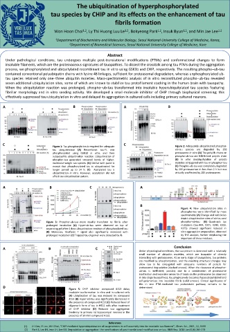
                          Figure 1: Tau phosphorylation is required for adequate   Figure 2: Adequately ubiquitinated phosphor-
                          tau  ubiquitination  (A)  Recombinat  tau-FL  was        ub-tau  species  are  degraded  by  26S
                          phosphorylated using GSK3β in vitro, and then            proteasome in vitro (A) The polyUb chains on
                          subjected to ubiquitination reaction. Ubiquitination of  phospho-ub-tau are K48-linked polyUb chain
                          phosphor-tau generated smeared bands of higher-          (B) In vitro deubiquitylation of polyUb
                          molecular-weight tau species. (B) Similar with panel A   moieties conjugated with tau or phosphor-tau
                          except that phosphorylated tau is ubiquitinated for      (C) Phospho–ub-tau was completely degraded
                          longer period up to 24 h. (C)  Acetylated tau is         by 26S proteasomes in less than 2 h but was
                          ubiquitination in vitro. However, acetylation did not    virtually unaffected by 20S proteasomes
                          affect tau ubiquitination patern.
  A                     B                  C                       A


                                                                   B
                                                                                      Figure 4: New ubiquitination sites in
                                                                                      phpospho-tau were identified by mass
                                                                                      spectrometry (A) Orange and red circles
                                                                                      depict ubiquitination sites of ub-tau and
                       Figure 3: Phosphor-ub-tau more readily transform to fibrils after  phosphor-ub-tau  (B) Quadruple Lys
                       prolonged incubation (A) Hyper-ub-tau were observed on top of  mutations (tau-4KR, K321, K343, K353,
                       separating gel after 4 days ubiquitination reaction of phosphorylated tau  K375) showed significant reduced in
                       (B) Moreover, thioflavin T signal also significantly increased with  vitro aggregation propensities, observed
                       prolonged incubation (C) Trapped tau oligomer were detected by IB.  by ThT analysis, further emphazing the
                                                                                      important of these residues.
  A                     B                     C                    Conclusion      Conclusion
                                                                  Under physiological conditions, the tau protein is decorated with a relatively
                                                                  small number of ubiquitin moieties, which are incapable of directly
                                                                  interacting with proteasomes. At an early stage of tauopathies, tau proteins
                                                                  are modified by phosphorylation, and the resulting structural changes may
                                                                  allow tau to be conjugated with adequate numbers of polyUb for
                                                                  proteasomal degradation (dashed arrows). When the clearance of phospho–
                                                                  ub-tau is inefficient, possibly due to a combination of proteasome
                                                                  inactivation and excessive amounts of loads on the proteasome (as observed
                                                                  in late-stage tauopathies), tau progressively becomes hyperubiquitylated and
                                                                  self-polymerizes into insoluble fibrils (solid arrows). Clinical significance of
                                                                  this in vivo PTM-mediated tau proteostatic pathway remains to be
  D                                                               determined.
                                 Figure 5: CHIP inhibitor compound #153 delay
                                 insoluble tau formation in vitro and in cultured cells
                                 (A) Ubiqitination of tau was reduced by compound
                                 #153 (B) Hyper-ub-tau was significantly decreased in
                                 the presence of compound #153 (C) Reduced level of
                                 oligomeric form of tau in HT22 cells after treatment
                                 of CHIP inhibitor (D) Reduced tau aggregation
                                 tendency in primary rat hippocampal neurons in the
                                 presence of 50 mM compound #153.
                                                    References
            (1)  JH Kim, JY Lee, WH Choi, “CHIP-mediated hyperubiquitination of tau promotes its self-assembly into the insoluble tau filament”, Chem. Sci., 2021, 12, 5599
             (2)  Park S, Lee JH, Jeon JH, Lee MJ. Degradation or aggregation: the ramifications of post-translational modifications on tau. BMB Rep. 2018 Jun;51(6):265-273

