
Page 3 - O. Microbiology
P. 3
The proliferation inhibitory effect of postbiotics made from antioxidizable
probiotics against HT-29 cell
2
1
Yeeun Kim and Keunho Ji *
1 Dept. of Microbiology, Pukyong Nat’l Univ. Busan, Korea
2 Basic Science Research Institute, Pukyong Nat’l Univ. Busan, Korea
BACKGROUND AIM
Postbiotics are the metabolites of probiotics, or the components that The primary goal of this study is to develop postbiotics that
result from probiotics activity in the gut, like fermentation compensate for the disadvantages of existing probiotics,
which are less resistant to the environment. Furthermore, it
proves the value of useful substances present in
extracellular and intracellular through the method of
manufacturing with postbiotics utilizing both the cells and
metabolites of cells. This study will be a study that proves
the usefulness of postbiotics.
METHODS RESULT
Preparation of postbiotics Table 1. Inhibition activity against HT-29 cell according to
Postbiotics was produced sonication-cell death method. concentration (ug/mL) of postbiotics
Overnight bacterial cultures were harvested by centrifugation
(3,500 g, 30 min, 4℃) and washed with 1X PBS buffer by conc 300 150 75 37.5 0
centrifugation (3,500 g, 30 min, 4℃). After centrifugation, Strain
discarded the supernatant and added 1X PBS buffer. Sonication La1 38.96 58.87 60.45 80.15 100
was performed under condition 20 rounds, 1 min/round, 70%
amplitude, 50W. After centrifugation, only the supernatant was La2 49.17 53.86 61.04 68.18 100
filtered and used in study.
Cell culture
In this study, human colon cancer cell line HT-29 cells and colon 50 La1
La2
normal cell line CCD-18Co cells were used. All cell lines were 40
purchased from Korean Cell Line Bank (Seoul, Korea). McCoy’s 30
5A medium containing 10% fetal bovine serum (FBS) was used Inhibition rate (%) 20
for HT-29 cells, and Dulbecco’s modified Eagle’s medium
(DMEM) containing 10% FBS was used for CCD-18Co cells. All 10
cell lines were cultured at 37℃, 5% CO conditions. After the 0 300 150 75 37.5 Control
2
Concentration (ug/mL)
cells were sufficiently adapted to the culture environment, when Figure 1. Anti-proliferation effect of postbiotics from La1 and La2.
the cell density was about 70~80% saturated, the cells were
passaged using 0.05% Trypsin-EDTA.
Cytotoxicity assay
The degree of inhibition of proliferation of HT-29 cells according
to the treatment by sample concentration was measured by
4-[3-(4-iodophenyl)-2-(4-nitrophenyl)-2H25-tetrazoliol]-1,3-
benzene disulphonate (WST-1) assay. WST-1 assay is a
method using WST-1 solution that is converted into a formazan,
a chromogen, by mitochondrial dehydrogenase of living cells,
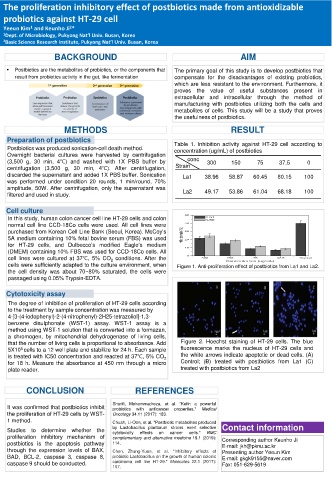
that the number of living cells is proportional to absorbance. Add Figure 2. Hoechst staining of HT-29 cells. The blue
5X10 cells to a 12 well plate and stabilize for 24 h. Each sample fluorescence marks the nucleus of HT-29 cells and
5
is treated with IC50 concentration and reacted at 37℃, 5% CO 2 the white arrows indicate apoptotic or dead cells. (A)
for 18 h. Measure the absorbance at 450 nm through a micro Control; (B) treated with postbiotics from La1 (C)
plate reader. treated with postbiotics from La2
CONCLUSION REFERENCES
It was confirmed that postbiotics inhibit Sharifi, Mohammadreza, et al. "Kefir: a powerful
anticancer
properties."
Medical
with
probiotics
the proliferation of HT-29 cells by WST- Oncology 34.11 (2017): 183.
1 method. Chuah, Li-Oon, et al. "Postbiotic metabolites produced
Studies to determine whether the by Lactobacillus plantarum strains exert selective Contact information
cells."
cytotoxicity
cancer
on
effects
BMC
proliferation inhibitory mechanism of complementary and alternative medicine 19.1 (2019): Corresponding author Keunho Ji
postbiotics is the apoptosis pathway 114. E-mail: jkh@pknu.ac.kr
through the expression levels of BAX, Chen, Zhung-Yuan, et al. "Inhibitory effects of Presenting author Yeeun Kim
BAD, BCL-2, caspase 3, caspase 8, probiotic Lactobacillus on the growth of human colonic E-mail: gkgk9155@naver.com
caspase 9 should be conducted. carcinoma cell line HT-29." Molecules 22.1 (2017): Fax: 051-629-5619
107.