
Page 29 - N. Metabolism and metabolic diseases
P. 29
Oxytocin-associated signaling is related with steroidogenesis and
differentiation of placenta
Sung-Min An¹, Min Jae Kim¹, Da Som Kim¹, So Young Kim¹, Da Hee Kang¹, Seung Chul Kim², Beum-Soo An¹ *
,
¹Department of Biomaterials Science, College of Natural Resources & Life Science/Life and Industry Convergence Research Institute, Pusan National University, Korea
2 Department of Obstetrics and Gynecology, Biomedical Research Institute, Pusan National University School of Medicine, Korea
Abstract Result
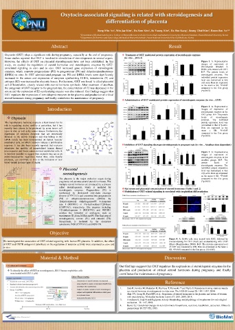
Oxytocin (OXT) plays a significant role during pregnancy, especially at the end of pregnancy. Treatment of OXT modulated protein expression of steroidogenic enzymes
Some studies reported that OXT is involved in stimulation of steroidogenesis in several organs. (in vitro _ JEG-3)
However, the effects of OXT on placental steroidogenesis have not been established. In this Figure 1. A: Representative
study, we studied the regulation of steroid hormones and steroidogenic enzymes by OXT- images of expression of in
steroidogenic
enzymes
associated signaling in vitro and in vivo. OXT increased gene expression of steroidogenic BeWo cells treated with OXT.
enzymes, which converts pregnenolone (PG) to progesterone (P4) and dehydroepiandrosterone B-F: The protein levels of
(DHEA) in vitro. In OXT administrated pregnant rat, PG and DHEA levels were significantly steroidogenic enzymes. The
increased in the serum and expression of enzymes synthesizing DHEA, testosterone (T) and individual protein expression
estrogen (E2) was increased in placenta tissues. Furthermore, OXT was found to affect placental level was normalized to that
of β-actin. Data are expressed
cell differentiation, closely related with steroid hormone synthesis. After treatment of atosiban, as the mean ± SD. *P<0.05
the antagonist of OXT receptor to the pregnant rats, the concentration of E2 was decreased in the compared to the Con group
serum and the expression of E2-synthesizing enzyme was also reduced. Our findings suggest that (*p<0.05)
OXT regulates the expression of steroidogenic enzymes in the placenta and production of critical
steroid hormones during pregnancy and finally contributes the maintenance of pregnancy.
Administration of OXT modulated protein expression of steroidogenic enzymes (in vivo _ OXT)
Introduction Figure 2. A: Representative
of
expression
images
of
steroidogenic enzymes in the
OXT group. B-F: The protein
Oxytocin levels of The steroidogenic
enzymes.
individual
The hypothalamic hormone oxytocin is best known for its protein expression level was
role in regulating uterine motility at parturition, but it has normalized to that of β-actin.
recently been shown to be produced by some tumor cell Data are expressed as the
types in vitro as well as by cancer tissues. Furthermore, the mean ± SD. *P<0.05
expression of oxytocin receptors that are structurally compared to the Con group
identical to the uterine receptors and that belong to the (*p<0.05)
GPCR family has been demonstrated on human vascular
endothelial cells, on which oxytocin induces a proliferative
response. It has also been recently reported that oxytocin Inhibition of OXT signaling decrease steroidogenesis in pregnant rats (in vivo _ Atosiban dose dependent)
stimulates the motility of immortalized human dermal
microvascular and breast cancer-derived endothelial cells. It Figure 3. A: Representative
is therefore possible to suggest that oxytocin may act as an images of expression of
endocrine/paracrine regulatory factor that, once locally steroidogenic enzymes in the
produced, can contribute in vivo to the formation of new atosiban groups. B-F: The
blood vessels in some types of cancer. protein levels of
steroidogenic enzymes. The
Placental individual protein expression
level was normalized to that
steroidogenesis of β-actin. Data are expressed
as the mean ± SD. *P<0.05
The placenta is the major endocrine organ during compared to the Con group
pregnancy and secretes several steroid hormones. The (*p<0.05)
multiple steroid hormones are produced by a process
called steroidogenesis, which is mediated by Rat serum and placental concentration of steroid hormones (Table 1 and 2)
steroidogenic enzymes. Pregnenolone (P5) is
synthesized by cholesterol side-chain cleavage Modulation of OXT-related signaling is correlated with trophoblast differentiation
enzyme (CYP11A1) and converted into progesterone
(P4) or dehydroepiandrosterone (DHEA) by
3β-hydroxysteroid dehydrogenase/δ5 4-isomerase
type 1 (HSD3B1) or 17α-hydroxylase/17,20-lyase
(CYP17A1), respectively. The enzymes including
17β-dehydrogenase 3 (HSD17B3) and HSD3B1
catalyze the formation of androgens, such as
testosterone (T), from DHEA and P5. The final step of
steroidogenesis, estrone (E1) and estradiol (E2)
biosynthesis, is mediated by the aromatase
cytochrome P450 (CYP19A1) and HSD17B.
Objective
Figure 4. A: BeWo cells were treated with OXT, followed by
We investigated the association of OXT-related signaling with human PE placenta. In addition, the effect immunostaining for CRH (Red) and counterstaining with DAPI
of OXT and OXTR antagonist (atosiban) on the regulation of invasion activity were examined in vitro and (Blue) (Magnification: 200X). B-D: The protein expression level
in vivo. of CRH was measured by WB. Data are expressed as the mean ±
SD. *P<0.05 compared to the Con group (*p<0.05)
Material & Method Discussion
Our findings suggest that OXT regulates the expression of steroidogenic enzymes in the
placenta and production of critical steroid hormones during pregnancy and finally
contributes the maintenance of pregnancy.
Other Experiments
• Concentration of steroid hormones was measured Reference
using competitive enzyme immunoassay (ELISA) kits,
according to the manufacturers
• Analysis of protein expression levels of steroidogenic 1. Sato K, Iemitsu M, Matsutani K, Kurihara T, Hamaoka T and Fujita S: Resistance training restores muscle
enzymes and trophoblast differentiation related genes sex steroid hormone steroidogenesis in older men. The FASEB Journal 28: 1891-1897, 2014.
in rat placenta tissue 2. Shin YY, Jeong JS, Park MN, et al.: Regulation of steroid hormones in the placenta and serum of women
• Immunocytochemistry (ICC) with preeclampsia. Molecular medicine reports 17: 2681-2688, 2018.
3. Furukawa S, Tsuji N and Sugiyama AJJotp: Morphology and physiology of rat placenta for toxicological
evaluation. 32: 1-17, 2019.
4. Buster JE: Gestational changes in steroid hormone biosynthesis, secretion, metabolism, and action. Clinics in
perinatology 10: 527-552, 1983.