
Page 33 - N. Metabolism and metabolic diseases
P. 33
Impact Of Heavy Metal Ion On Cartilage Degeneration
2
1
Godagama Gamaarachchige Dinesh Suminda , Dahye Kim , Yunhui Min , Xiangyu Zhao , Mangeun Kim , Young-Ok Son 1,2*
1
2
1
1 Interdisciplinary Graduate Program in Advanced Convergence Technology and Science, Jeju National University, Jeju Special Self-Governing Province, 63243, Republic of Korea.
2 Department of Animal Biotechnology, Faculty of Biotechnology, College of Applied Life Sciences, Jeju National University, Jeju Special Self-Governing Province, 63243, Republic of Korea.
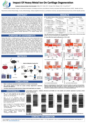
ABSTRACT RESULTS
Osteoarthritis (OA) is the whole-joint disease and the most common form of arthritis. The A. The viability effects of Heavy metal Ion in primary culture articular chondrocytes
imbalance of anabolic and catabolic factors was identified as the main cause of OA. Metal ion
pollution is a serious environmental problem worldwide, Therefore, heavy metal can expose to
the human body through drinking water, food, and inhalation caused many serious diseases.
However, no reports addressing the role of the heavy metal ion in OA progression. Here, we
investigated the main heavy metals ions (Cd, Cr, As, Ni) effect on OA pathogenesis. First, we
evaluated the cytotoxic effects of Cd (0 - 1 µM), Cr (0 - 1 µM), As (0 - 10 µM), and Ni (0- 1 mM)
by the MTT assay. RT-PCR results revealed that Cd, Cr, As, and Ni ion increase the
metalloproteinases (MMP2, MMP3, MMP9, MMP10, MMP12, and MMP13), ZIP8, and Epas1
expression significantly in articular chondrocytes. Further, the cartilage degeneration was
shown in mice joints by intra-articular injection of heavy metals. The low-dose, long-term oral
administration of heavy metal by drinking experiments are ongoing. In conclusion, our findings
suggest that metal ion can influence cartilage degeneration in the articular cartilage, which
could affect OA development.
Fig. 2. MTT assay results confirming the in vitro cytotoxicity effect of heavy metal ion (Cd2+/Cr2+/As3+/Ni2+)
PURPOSE OF EXPERIMENTS against the articular chondrocytes for 48 h (unpublished data).
B. Heavy Metal Ion regulate pathogenesis of OA in mice
Osteoarthritis (OA) is a leading cause of disability and bears a large socioeconomic cost.
Cartilage destruction, synovial inflammation, osteophyte formation, and subchondral bone
sclerosis are the well-known symptoms of OA pathogenesis. OA is caused by imbalance of an
anabolic and catabolic factors that is induced by a variety of etiologic risk factors and
pathophysiological processes, including mechanical injury, genetic factors, aging, obesity,
gender, and metabolic disorder. These factors alter biochemical mechanisms in chondrocytes,
resulting in degradation of the extracellular matrix (ECM). However, no Sufficient data about
Environmental factors and particular reason for OA disease has been investigated to date.
Metal ion pollution is a serious environmental problem worldwide, heavy metal can expose to
the human body through drinking water, food, and inhalation caused many serious diseases.
China groundwater arsenic level over the limit of 10 μg/L for drinking water. It is worth
continuously paying attention to metal ion exposure related public health problems. Here, we
used the influence of metal ion and approaches to investigate whether the metal ions are
associated with OA pathogenesis.
METHODS
CONCLUSION
Metal ion enhanced catabolic factors expression in articular chondrocytes.
Fig.3.(a,b,c,d) Experimental OA was examined by safranin-O staining and scoring of OA parameters,
Intra articular injection of metal ion caused cartilage degeneration especially, including cartilage destruction (OARSI grade), synovitis, osteophyte maturity and thickening of the
chromium and Cadmium. subchondral bone plate. Safranin-O staining and scoring of OA parameters in wild-type mice that had
Low-dose, long-term oral administration of heavy metal experiments are ongoing. undergone intra-articular injection of heavy metal ion for 3 weeks(unpublished data)
REFERENCES C. The effects of Arsenic , Cadmium, Chromium and Nickle Ion in the anabolic and catabolic expression in primary culture
articular chondrocytes.
• Kim, J. H. Et Al. Regulation Of The Catabolic
Cascade In Osteoarthritis By The Zinc-zip8-mtf1
Axis. Cell 156, 730-743 (2014).
• Won, Y. Et Al. Pleiotropic Roles Of
Metallothioneins As Regulators Of Chondrocyte
Apoptosis And Catabolic And Anabolic Pathways
During Osteoarthritis Pathogenesis. Ann. Rheum.
Dis.75, 2045-2052 (2016).
• Bauer, Christoph, Et Al. "Concentration-dependent
Effects Of Cobalt And Chromium Ions On
Osteoarthritic Chondrocytes." Cartilage (2019):
1947603519889389.
• Chung, Yao-pang, Et Al. "Arsenic Induces Human
Chondrocyte Senescence And Accelerates Rat
Articular Cartilage Aging." Archives Of
Toxicology 94.1 (2020): 89-101.
Fig. 1. Effect of gene activity in chondrocytes treated with 0μM,0.1μM,0.2μM,0.5μM of metal Ion concertation (a) Anabolic factors (b) Catabolic Factors (c)
Metrix Metalloproteinase(unpublished data)
CONTACT INFORMATION
Name-Godagama Gamaarachchige Dinesh Suminda, Sustainable Agriculture Research Institute 111, 102, Jejudaehak-ro, Jeju-si, Jeju Special Self-Governing Province, 63243 Republic of
Korea, E-mail: dsuminda00@gmail.com