
Page 35 - N. Metabolism and metabolic diseases
P. 35
The role of pentose phosphate pathway in pulmonary fibrosisThe role of pentose phosphate pathway in pulmonary fibrosis
Seung Hyun Kwon, Hak Su Kim
S e u n g H y u n K w o n , H a k S u K i m
Veterans Medical Research Institute, Veterans Health Service Medical Center, Seoul, Republic of Korea
V e t e r a n s M e d i c a l R e s e a r c h I n s t i t u t e , V e t e r a n s H e a l t h S e r v i c e M e d i c a l C e n t e r , S e o u l , R e p u b l i c o f K o r e a
BACKGROUND AIM
Pentose phosphate (PP) pathway is one of the major metabolic pathways associated with glucose This study aims to investigate the
metabolism. Glucose 6-phosphate dehydrogenase (G6PD) is a rate-limiting enzyme of PP pathway role of the PP pathway in
and its function is inhibited by 6-aminonicotinamide (6-AN). However, the role of PP pathway in lung idiopathic pulmonary fibrosis (IPF).
fibrosis remain unexplored.
METHODS
The metabolites of PP pathway were analyzed with Agilent 7890/5975 GC/MSD system and HP-5 MS column in the human lung tissues (IPF
=31 and control=20). The roles of G6PD were evaluated using fibrotic markers in fibroblasts or epithelial cells treated with transforming
growth factor-β1 (TGF-β1). The antifibrotic role of 6-AN was assessed in a bleomycin-induced lung fibrosis mice model. The levels of
proteins in cell lysates or tissues were measured by western blot assays or RT-PCR.
RESULTS
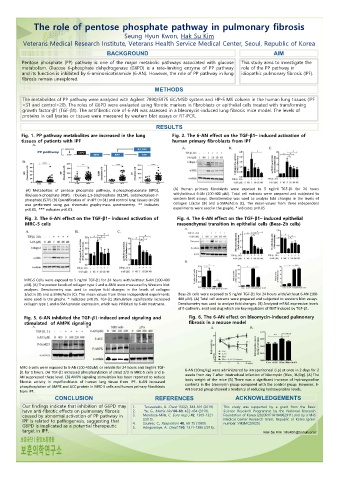
Fig. 1. PP pathway metabolites are increased in the lung Fig. 2. The 6-AN effect on the TGF-β1– induced activation of
tissues of patients with IPF human primary fibroblasts from IPF
(A) Metabolites of pentose phosphate pathway, 6-phosphogluconate (6PG), (A) Human primary fibroblasts were exposed to 5 ng/ml TGF-β1 for 24 hours
ribulose-5-phosphate (R5P), ribulose-1,5-bisphosphate (R1,5P), sedoheptulose-7- with/without 6-AN (100-400 μM). Total cell extracts were prepared and subjected to
phosphate (S7P) (B) Quantification of in IPF (n=31) and control lung tissues (n=20) western blot assays. Densitometry was used to analyze fold changes in the levels of
collagen 1/actin (B) and α-SMA/Actin (C). The mean-values from three independent
was performed using gas chromato graphy-mass spectrometry. ** indicates
p<0.01, *** indicates p<0.01. experiments were used in the graphs. * indicates p<0.05
Fig. 3. The 6-AN effect on the TGF-β1– induced activation of Fig. 4. The 6-AN effect on the TGF-β1– induced epithelial
MRC-5 cells mesenchymal transition in epithelial cells (Beas-2b cells)
MRC-5 Cells were exposed to 5 ng/ml TGF-β1 for 24 hours with/without 6-AN (100-400
μM). (A) The protein levels of collagen type 1 and α-SMA were measured by Western blot
analyses. Densitometry was used to analyze fold changes in the levels of collagen
1/actin (B) and α-SMA/Actin (C). The mean values from three independent experiments Beas-2b cells were exposed to 5 ng/ml TGF-β1 for 24 hours with/without 6-AN (100-
were used in the graphs. * indicates p<0.05. TGF-β1 stimulation significantly increased 400 μM). (A) Total cell extracts were prepared and subjected to western blot assays.
collagen type 1 and α-SMA protein expression, which was inhibited by 6-AN treatment. Densitometry was used to analyze fold changes. (B) Analyzed mRNA expression levels
of E-cadherin, snail and slug which are key regulators of EMT induced by TGF-β1.
Fig. 5. 6-AN inhibited the TGF--induced smad signaling and Fig. 6. The 6-AN effect on bleomycin-induced pulmonary
stimulated of AMPK signaling fibrosis in a mouse model
MRC-5 cells were exposed to 6-AN (100-400uM) or vehicle for 24 hours and 5ng/ml TGF- 6-AN (10mg/kg) were administered by intraperitoneal (i.p) at once in 2 days for 2
β1 for 1 hours. (A) TGF-β1 increased phosphorylation of smad 2/3 in MRC-5 cells and 6-
AN suppressed these level. (B) AMPK signaling stimulation has been reported to reduce weeks from day 7 after intatracheal infection of bleomycin (Bleo, 3U/kg). (A) The
body weight of the mice (B) There was a significant increase of hydroxyproline
fibrotic activity in myofibroblasts of human lung tissue from IPF. 6-AN increased
phosphorylation of AMPK and ACC protein in MRC-5 cells and human primary fibroblasts contents in the bleomycin group compared with the control group. However, 6-
AN treated group showed a tendency of reducing hydroxyproline levels.
from IPF.
CONCLUSION REFERENCES ACKNOWLEDGEMENTS
Our findings indicate that inhibition of G6PD may 1. Tzouvelekis, A. Chest 156(2), 383-391 (2019). This study was supported by a grant from the Basic
have anti-fibrotic effects on pulmonary fibrosis 2. Yu, G., Matrix biol 68-69, 422-434 (2018). Science Research Programme by the National Research
caused by abnormal activation of PP pathway in 3. Mendoza-Milla, C. Euro resp j 42, 1309-1321 Foundation of Korea (2020R1F1A104962911) and by a VHS
IPF is related to pathogenesis, suggesting that 4. (2013). Medical Center Research Grant, Republic of Korea (grant
number: VHSMC20025)
Saunier, C., Respiration 40, 69-75 (1980).
G6PD is implicated as a potential therapeutic 5. Adegunsoye, A. Chest 150, 1371-1386 (2016).
target in IPF. Hak Su Kim : khs401@bohun.or.kr