
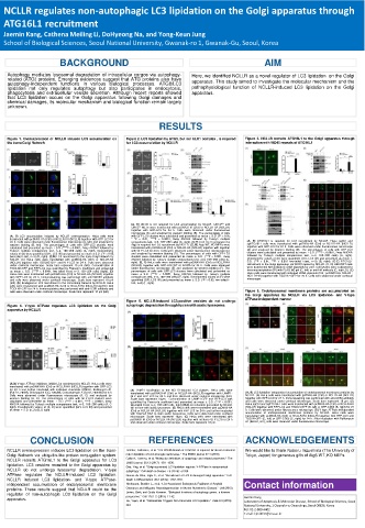
Page 27 - F. Cell biology
P. 27
NCLLR regulates non-autophagic LC3 lipidation on the Golgi apparatus through
  ATG16L1 recruitment
  Jaemin Kang, Cathena Meiling Li, DoHyeong Na, and Yong-Keun Jung
  School of Biological Sciences, Seoul National University, Gwanak-ro 1, Gwanak-Gu, Seoul, Korea

                   BACKGROUND                                                  AIM

   Autophagy mediates lysosomal degradation of intracellular cargos via autophagy-  Here, we identified NCLLR as a novel regulator of LC3 lipidation on the Golgi
   related (ATG) proteins. Emerging evidences suggest that ATG proteins also have
   autophagy-independent functions in various biological processes. ATG8/LC3  apparatus. This study aimed to investigate the molecular mechanism and the
   lipidation not only regulates autophagy but also participates in endocytosis,  pathophysiological function of NCLLR-induced LC3 lipidation on the Golgi
   phagocytosis and extracellular vesicle secretion. Although recent reports showed  apparatus.
   that LC3 lipidation occurs on the Golgi apparatus following Golgi damages and
   chemical damages, its molecular mechanism and biological function remain largely
   unknown.
                                                RESULTS

   Figure 1. Overexpression of NCLLR induces LC3 accumulation on  Figure 2. LC3 lipidation by ATG5, but not ULK1 complex , is required  Figure 3. NCLLR recruits ATG16L1 to the Golgi apparatus through
   the trans-Golgi Network            for LC3 accumulation by NCLLR       interaction with WD40 repeats of ATG16L1


                                        (A, B) Ulk1/2 is not required for LC3 accumulation by NCLLR. Ulk1/2 +/+ and
                                        Ulk1/2 -/- MEFs were transfected with pcDNA3-HA (Ctrl) or NCLLR-HA (NCLLR)
                                        together with GFP-LC3 for 24 h. Cells were observed under fluorescence
                                        microscope (A) and analyzed by western blotting (B). The percentages of cells
    (A, B) LC3 accumulation induced by NCLLR overexpression. HeLa cells were  with GFP-LC3 clusters were calculated and presented as mean ± S.D. (*P < 0.05,
    transfected with pcDNA3-HA (Ctrl) or NCLLR-HA (NCLLR) together with GFP-LC3 for  * *P < 0.01, ****P < 0.0001, 2way ANOVA followed by Tukey’s multiple
    24 h. Cells were observed under fluorescence microscope (A, left) and analyzed by  comparisons test, n=3, 100~250 cells) (A, right). (C-F) LC3 I-to-II conversion by  (A, B) ATG16L1 is required for LC3 recruitment by NCLLR. HeLa sgCtrl and
                                                                          sgATG16L1 cells were transfected with pcDNA3-HA (Ctrl) or NCLLR-HA (NCLLR)
    western blotting (B, left). The percentages of cells with GFP-LC3 clusters were  Atg5 is required for LC3 recruitment by NCLLR. (C, D) Atg5 WT, KO MEFs were
    calculated and presented as mean ± S.D. (****P < 0.0001, 2way ANOVA followed by  transfected with pcDNA3-HA (Ctrl) or NCLLR-HA (NCLLR) together with together  together with GFP-LC3 for 24 h. Cells were observed under fluorescence microscope
    Tukey’s multiple comparisons test, n=3, 100~200 cells) (A, right). Densitometry  with GFP-LC3 for 24 h. Cells were observed under fluorescence microscope (C)  (A) and analyzed by western blotting (B). The percentages of cells with GFP-LC3
                                                                          clusters were calculated and presented as mean ± S.D. (****P < 0.0001, 2way ANOVA
    values of (B) were quantified (GFL-LC3 II/I) and presented as mean ± S.D. (*P < 0.05,  and analyzed by western blotting (D). The percentages of cells with GFP-LC3
    two-tailed t-test, n=3) (B, right). (C,D) LC3 recruitment to the trans-Golgi Network by  clusters were calculated and presented as mean ± S.D. (***P < 0.001, 2way  followed by Tukey’s multiple comparisons test, n=3, 100~200 cells) (A, right).
    NCLLR. (C) HeLa cells were transfected with pcDNA3-HA (Ctrl) or NCLLR-HA  ANOVA followed by Tukey’s multiple comparisons test, n=3, 100~250 cells) (C,  Densitometry values of (B) were quantified (GFL-LC3 II/I) and presented as mean ±
                                                                          S.D. (**P < 0.01, ***P < 0.001 two-tailed t-test, n=3) (B, right). (C-E) ATG16L1
    (NCLLR) together with TGOLN2-GFP and RFP-LC3 for 24 h. Cells were observed  right). (E, F) HeLa cells were transfected with pcDNA3-HA (Ctrl) or NCLLR-HA  recruitment to the Golgi apparatus via WD40 repeat by NCLLR. (C, D) HEK293T cells
    under confocal microscope. Scale bars represent 10 μm (left). Co-localization of  (NCLLR) together with GFP-LC3 WT or G120A for 24 h. Cells were observed
    TGOLN2-GFP and RFP-LC3 was quantified by Pearson’s coefficient and presented  under fluorescence microscope (E) and analyzed by western blotting (F). The  were transfected with indicated cDNA plasmids for 24 h. Cell lysates were subjected to
                                                                          immunoprecipitation (IP) with FLAG M2 gel (C, left) or anti-HA antibody (C, right, D). (E)
    as mean ± S.D. (****P < 0.0001, two-tailed t-test, n=3, 150~250 cells) (right). (D)  percentages of cells with GFP-LC3 clusters were calculated and presented as  HeLa cells were transfected with indicated cDNA plasmids (Ctrl : pcDNA3-HA, NCLLR :
    HeLa cells were transfected with pcDNA3-HA (Ctrl) or NCLLR-HA (NCLLR) together  mean ± S.D. (****P < 0.0001, 2way ANOVA followed by Tukey’s multiple
    with GFP-LC3 for 24 h. Immunostaining was performed with anti-GM130 antibody  comparisons test, n=3, 100~200 cells) (E, right). Densitometry values of (F) were  NCLLR-HA) together with TGOLN2-GFP for 24 h. Cells were observed under confocal
                                                                          microscope.
    and cells were observed under confocal microscope. Scale bars represent 10 μm  quantified (GFL-LC3 II/I) and presented as mean ± S.D. (*P < 0.05, two-tailed t-
    (left). (E) Endogenous LC3 recruitment to the trans-Golgi Network by NCLLR. HeLa  test, n=3) (F, right).
    cells were transfected with pcDNA3-HA (Ctrl) or NCLLR-HA (NCLLR) together with
    TGOLN2-GFP for 24 h. Immunostaining was performed with anti-LC3B antibody and  Figure 6. Endolysosomal membrane proteins are accumulated on
    cells were observed under confocal microscope. Scale bars represent 10 μm (left).
                                                                          the Golgi apparatus by NCLLR via LC3 lipidation- and V-type
                                                                          ATPase-independent manner
                                      Figure 5. NCLLR-induced LC3-positive vesicles do not undergo
   Figure 4. V-type ATPase regulates LC3 lipidation on the Golgi  autophagic degradation through fusion with acidic lysosomes
   apparatus by NCLLR
    (A-D) V-type ATPase inhibitors inhibit LC3 recruitment by NCLLR. HeLa cells were
    transfected with pcDNA3-HA (Ctrl) or NCLLR-HA (NCLLR) together with GFP-LC3
    for 24 h and further incubated with indicated chemicals (DMSO, Bafilomycin A1  (A) LAMP1 localization to the NCLLR-induced LC3 clusters. HeLa cells were
    (Baf.A1, 20nM), Chloroquine (CQ, 100uM), Concanamycin A (ConA, 200nM)) for 6 h.  transfected with pcDNA3-HA (Ctrl) or NCLLR-HA (NCLLR) together with LAMP1-  (A, B) LC3 lipidation-independent accumulation of endolysosomal membrane proteins by
    Cells were observed under fluorescence microscope (A, C) and analyzed by  GFP and RFP-LC3 for 24 h and then observed under confocal microscope (left).  NCLLR. (A) HeLa cells were transfected with pcDNA3-HA (Ctrl) or NCLLR-HA (NCLLR)
    western blotting (B, D). The percentages of cells with GFP-LC3 clusters were  Scale bars represent 10μm.  Co-localization of LAMP1-GFP and RFP-LC3 was  together with GFP-LC3 for 24 h. Immunostaining was performed with anti-CD63 antibody
    calculated and presented as mean ± S.D. (***P < 0.001, ****P < 0.0001, 2way  quantified by Pearson’s coefficient and presented as mean ± S.D. (***P < 0.001,  and cells were observed under confocal microscope. Scale bars represent 10 μm. (B)
    ANOVA followed by Tukey’s multiple comparisons test, n=3, 100~200 cells) (A, C  two-tailed t test, n=3, 100~200 cells, right) (B,C) LC3 clusters generated by NCLLR  HeLa sgCtrl and sgATG16L1 cells were transfected with pcDNA3-HA (Ctrl) or NCLLR-HA
    right). Densitometry values of (B, D) were quantified (GFL-LC3 II/I) and presented  fail to fuse with acidic lysosomes. (B) HeLa cells were transfected with pcDNA3-HA  (NCLLR) together with RFP-LC3 and TGOLN-GFP (B, left) or GFP-CD63 (B, right) for 24
    as mean ± S.D. (n=3) (B, D right)   (Ctrl) or NCLLR-HA (NCLLR) together with RFP-LC3 for 24 h and further incubated  h. Cells were observed under fluorescence microscope. (C) V-type ATPase-independent
                                        with DQ-Red BSA to stain acidic lysosomes. Cells were observed under confocal  accumulation of endolysosomal membrane proteins by NCLLR. HeLa cells were
                                        microscope. Scale bars represent 10μm. (C) HeLa cells were transfected with  transfected with pcDNA3-HA (Ctrl) or NCLLR-HA (NCLLR) together with RFP-LC3 and
                                        pcDNA3-HA (Ctrl) or NCLLR-HA (NCLLR) together with mCherry-GFP-LC3 for 24 h  TGOLN-GFP (C, left) or GFP-CD63 (C, right) for 24 h. After incubation with Bafilomycin
                                        and observed under confocal microscope. Scale bars represent 10 μm.  A1 (Baf.A1, 6 h), cells were observed under fluorescence microscope.
          CONCLUSION                         REFERENCES                   ACKNOWLEDGEMENTS
   NCLLR overexpression induces LC3 lipidation on the trans-  Fletcher,  Katherine, et al. "The WD40 domain of ATG16L1 is required  for its non‐canonical   We would like to thank Noboru mizushima (The University of
   Golgi Network via ubiquitin-like protein conjugation system.  role in lipidation of LC3 at single membranes." The EMBO journal 37.4 (2018).  Tokyo, Japan) for generous gifts of Atg5 WT, KO MEFs.
   NCLLR recruits ATG16L1 to the Golgi apparatus for LC3  Galluzzi, Lorenzo, et al. "Molecular definitions of autophagy  and related  processes." The
   lipidation. LC3 vesicles recruited to the Golgi apparatus by  EMBO journal  36.13 (2017): 1811-1836.
                                      Gao, Ying, et al. "Golgi-associated LC3 lipidation requires V-ATPase in noncanonical
   NCLLR do not undergo lysosomal degradation. V-type  autophagy." Cell death & disease 7.8 (2016): e2330.
   ATPase regulates the NCLLR-induced LC3 lipidation.  Gomes-da-Silva, Lígia C., et al. "Recruitment of LC3 to damaged Golgi apparatus." Cell
   NCLLR induced LC3 lipidation- and V-type ATPase-  Death & Differentiation  26.8 (2019): 1467-1484.
   independent accumulation of endolysosomal membrane  Heckmann, Bradlee L., et al. "LC3-Associated  Endocytosis Facilitates β-Amyloid   Contact information
   proteins. These results suggest that NCLLR could be the  Clearance and Mitigates Neurodegeneration  in Murine  Alzheimer’s Disease." Cell (2019).
   regulator of non-autophagic LC3 lipidation on the Golgi  Levine,  Beth, and Guido Kroemer.  "Biological functions of autophagy genes:  a disease
   apparatus.                         perspective." Cell 176.1-2 (2019): 11-42.  Jaemin Kang
                                                                         Laboratory of Apoptosis & Molecular Disease, School of Biological Sciences, Seoul
                                      Liu, Yajun, et al. "Niclosamide Triggers Non-Canonical  LC3 Lipidation." Cells 8.3 (2019):
                                      248.                               National University, 1 Gwanak-ro, Gwanak-gu, Seoul 08826, Korea
                                                                         Tel: 82-2-880-4402
                                                                         E-mail: kjm0831@snu.ac.kr