
Page 23 - F. Cell biology
P. 23
Dual regulation of apoptosis and autophagy by the target in
chloroquine-treated ARPE-19 cells
1
1
Anh-Thu Nguyen-Hoang ; Hoang-Long Ngo ; Sook-Jeong Lee 1
1 Department of Bioactive Material Science, Jeonbuk National University, Jeonju, South Korea
BACKGROUND AIM
Chloroquine (CQ), a compound of 4-aminoquinoine, has been commonly used as The serine/threonine protein kinases play an important role in
an antimalarial and anti-inflammatory drug. Currently, it is widely prescribed for eukaryotic signaling pathways, such as cell differentiation,
treatment of amebiasis, rheumatoid arthritis, systemic lupus erythematosus, and proliferation, and cell metabolisms. Here we experiment to
for prophylaxis against malaria. However, retinal toxicity with the retinal pigment elucidate the role of one of the inhibitors for the serine/threonine
epithelial (RPE) degeneration and neurosensory retina as a result of long-term protein kinases on CQ-induced RPE toxicity.
routine use of CQ has been well defined, as CQ were examined to cause vacuoles
formation and cell death in human retinal pigment epithelium-derived cells.
METHODS
Cell Culture The ARPE-19, a spontaneously immortalized cell line of human retinal pigment epithelium, obtained from ATCC (Manassas, VA,
USA) was maintained in Dulbecco’s Modified Eagle’s medium (DMEM) and Ham’s F-12 media containing 10% fetal bovine serum and 1% (v/v)
penicillin/streptomycin.
MTT assay To determine cytotoxicity after combined treatment with CQ and a target inhibitor in ARPE-19 cells
Western blot To identify altered signal protein expressions associated with the CQ and a target inhibitor treatment, cell lysates extracted were
performed the western blotting analysis.
FACS analysis To analyze apoptotic cell death, ARPE-19 cells treated with different combinations of drugs stained with Annexin V/PI stains.
RESULTS
Figure 1. Target inhibitor effect on CQ-induced vacuole formation and
cell death in ARPE-19 cells
Attenuated CQ cytotoxicity and vacuole
formation by the target inhibitor (A)
(B) 150
Fig. 1. Target inhibitor effect on vacuole CQ
CQ + T-inhibitor, 10M
formation and cell death in ARPE-19 cells ns ns
APRE-19 cells were treated with various concentrations of 100 * ** *** **
CQ (0 to 120 μM) alone or together with 10 μM target % Survival
inhibitor for 24 h. (A) Phase-contrast photomicrographs of 50
ARPE-19 cells treated with individual drugs. (B) Percent
survival of ARPE-19 cells. Bars indicates relative survival
changes (mean SD, n=3, ns; not significant, *P<0.05, 0
**P<0.01, ***P<0.001 vs. CQ alone-treated each control). 0 10 50 75 100 120
CQ [M]
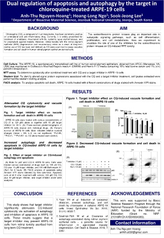
Increased autophagy and decreased Figure 2. Decreased CQ-induced vacuole formation and cell death in
apoptosis in CQ-treated ARPE-19 cells by ARPE-19 cells
target inhibitor
(B)
Fig. 2. Effect of target inhibitor on CQ-induced
autophagy and apoptosis (A)
(A) Blots for p62 and LC3 in ARPE-19 cells. Cells were
treated various combination of drugs such as 100 μM CQ,
CQ plus 10 μM target inhibitor, and 100 nM Bafilomycin A1
(Ba1) for 6 hours. (B) FACS analysis of apoptosis using
Annexin V/PI stains detected by flow cytometer. Apoptotic
cells at 24 h after treatment with vehicle, 100 μM CQ, CQ
plus 10 μM target inhibitor, and target inhibitor alone were
analyzed.
CONCLUSION REFERENCES ACKNOWLEDGEMENTS
1. Yoon YH et al. Induction of lysosomal This work was supported by Basic
dilatation, arrested autophagy, and cell
This study shows that target inhibitor death by chloroquine in cultured ARPE-19 Science Research Program through the
significantly attenuates CQ-induced cells. Invest Ophthalmol Vis Sci. 2010; National Research Foundation of Korea
cytotoxicity via activation of autophagy 51(11): 6030-7. (NRF) funded by the Ministry of
and inhibition of apoptosis in ARPE-19 Education (Grant no. NRF-
cells. These results suggest that a 2. Szatmári-Tóth M et al. Clearance of 21016R1D1A1B04934383).
autophagy-associated dying retinal pigment
target inhibitor may prevent ARPE-19 epithelial cells – a possible source for Contact information
cells from retinal toxicity ascribed from inflammation in age-related macular
long term CQ treatment. degeneration. Cell Death & Disease. 2016; 7: Anh-Thu Nguyen-Hoang:
2367.
anhthu232@gmail.com