
Page 21 - F. Cell biology
P. 21
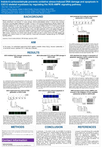
Indole-6-carboxaldehyde prevents oxidative stress-induced DNA damage and apoptosis in
  C2C12 skeletal myoblasts by regulating the ROS-AMPK signaling pathway
                     2,3
         1
   Cheol Park , Yung Hyun Choi *
  1 Division of Basic Sciences, College of Liberal Studies, Dong‐eui University, Busan 47340,
  2 Department of Biochemistry, Dong-eui University College of Korean Medicine, Busan 47227,
  3 Anti-Aging Research Center, Dong-eui University, Busan 47340, Republic of Korea
                          BACKGROUND                                       I6CA protected H 2 O 2 -induced mitochondrial
                                                                                 dysfunction in C2C12 cells
    Myoblast damage due to oxidative stress is one of the main causes of skeletal muscle loss associated with the inhibition of
    myopic differentiation and induction of muscle damage. Indole-6-carboxaldehyde (I6CA), a natural indole derivative
    derived from the brown algae Sargassum thunbergii, is known to have several pharmacological activities. However, the
    antioxidative effects of I6CA have not been identified. In this study, we investigated that the protective effect of I6CA and its
    underlying mechanism in vitro using hydrogen peroxide (H 2 O 2 )-induced oxidative stress in a C2C12 mouse skeletal
    myoblast. The findings revealed that pretreatment with I6CA protected H 2 O 2 -induced cytotoxicity and DNA damage by
    blockage of ROS generation. Further studies have shown that I6CA suppressed C2C12 cells against H 2 O 2 -induced
    apoptosis by preventing loss of mitochondrial membrane potential. I6CA attenuated H 2 O 2 -induced activation of AMPK and
    ATP content. Additionally, the cytoprotective effects of I6CA against H 2 O 2 were eliminated by compound C, a specific
    AMPK blocker. Collectively, the current results indicate that I6CA was able to protect C2C12 cells DNA damage and
    apoptosis from oxidative stress by at least preserving mitochondrial homeostasis mediated through the ROS-AMPK
    signaling pathway.
    Keywords: Indole 6 carboxaldehyde; DNA damage; apoptosis; AMPK
                                  AIM
                                                                          Fig. 5. Inhibition of H 2 O 2 -induced mitochondrial dysfunction by I6CA in C2C12 cells.
                                                                          The cells were treated with 400 μM I6CA or 10 mM NAC for 1 h and then exposed to 1
                                                                          mM H 2 O 2 for 24 h. (A) The cells were collected and stained with JC-1. The JC-1
    In this study, the antioxidant potential of I6CA against oxidative stress (H 2 O 2 )-induced cytotoxicity in  fluorescence intensity was detected to evaluate the changes in the MMP using a flow
                                                                          cytometer. (B) The percentage of cells with JC-1 monomers is indicated by bars, and
    immortalized mouse myoblast C2C12 cells was evaluated.                the data represent the mean ± SD of triplicate determinations (*** p< 0.001 compared
                                                                          with the control group; ### p< 0.001 compared with the H 2 O 2 -treated group). (C) JC-1
                                                                          fluorescence images of the cells treated with 1 mM H 2 O 2 in the presence or absence of
                                                                          400 μM I6CA are shown. Red fluorescence indicates high membrane potential, and
                              RESULTS                                     green fluorescence represents low membrane potential. Representative images were
                                                                          captured using a fluorescence microscope (original magnification, ×400).
      I6CA inhibited H 2 O 2 -induced cytotoxicity in   I6CA attenuated H 2 O 2 -induced DNA damage in   I6CA abolished the change in expression of
                C2C12 cells                        C2C12 cells             apoptosis regulatory factors caused by H 2 O 2
                                                                                     in C2C12 cells


                                                                          Fig. 6. Effects of I6CA on the expression of apoptosis regulators in H 2 O 2 -treated
                                                                          C2C12 cells. The cells were treated with or without 400 μM I6CA for 1 h before
                                       Fig. 3. Protection of H 2 O 2 -induced DNA damage by I6CA in C2C12 cells. The cells  treatment with 1 mM H 2 O 2 for 24 h. (A) Cytochrome c levels were analyzed by Western
    Fig. 1. Protective effect of I6CA on H 2 O 2 -induced cytotoxicity in C2C12 cells. The cells  were treated with or without 400 μM I6CA for 1 h before treatment with 1 mM H 2 O 2 for  blotting on mitochondrial and cytoplasmic fractions isolated from cells. Cytochrome
    were treated with the various concentrations of I6CA for 24 h (A), or pretreated with or  24 h. (A) A comet assay was performed, and representative images were captured  oxidase subunit VI (COX IV) and actin serve as protein loading controls for the
    without the indicated concentrations of I6CA or 10 mM NAC for 1 h, and then cultured  using a fluorescence microscope (original magnification, ×200). (B) The cell lysates  mitochondria and cytosol, respectively. (B) Whole cell lysates were prepared, and Bax,
    in the presence of 1 mM H 2 O 2 for 24 h (B and C). (A and B) The cell viability was  were prepared, and p-γH2AX and γH2AX expression was identified by Western blot  Bcl-2, caspase-9, caspase-3 and PARP expressions were identified by Western blot
    determined by an MTT reduction assay. The results are expressed as the mean ± SD  analysis. The equivalent loading of proteins in each well was confirmed by actin. (C)  analysis. The equivalent loading of proteins in each well was confirmed by actin. (C
    obtained from three independent experiments (* p < 0.05, and *** p < 0.001 compared  The DNA samples of cells were subjected to assessment of the 8-OHdG levels. The  and D) The activities of caspase-9 (C) and caspase-3 (D) in cell lysates were
    with the control group; # p < 0.05 ### p < 0.01 and ### p < 0.001 compared with the  measurements were made in triplicate, and the results are expressed as the mean ±  measured using the respective substrate peptides. The measurements were made in
    H 2 O 2 -treated group). (C) Representative images of the cells were captured by a phase-  SD (*** p < 0.001 compared with the control group; ## p < 0.01 compared with the  triplicate, and the results are expressed as the mean ± SD (*** p< 0.001 compared
    contrast microscope (original magnification, 200×).  H 2 O 2 -treated group).  with the control group; ### p< 0.001 compared with the H 2 O 2 -treated group).
        I6CA suppressed H 2 O 2 -induced ROS   I6CA reduced H 2 O 2 -induced apoptosis in   AMP-activated protein kinase (AMPK) was
           generation in C2C12 cells               C2C12 cells             involved in the mitigation of H 2 O 2 -mediated
                                                                              cytotoxicity by I6CA in C2C12 cells


                                       Fig. 4. Inhibitory effect of I6CA on H 2 O 2 -induced apoptosis in C2C12 cells. The cells
                                       were treated with or without 200 or 400 μM I6CA for 1 h before treatment with 1 mM
                                       H 2 O 2 for 24 h. (A) The cells were stained with DAPI solution and stained nuclei were
                                       observed using a fluorescence microscope (original magnification, ×200). Each image
    Fig. 2. Attenuation of H 2 O 2 -induced ROS generation by I6CA in C2C12 cells. The cells  is representative of at least three independent experiments. (B and C) The cells were  Fig. 7. The relevance of the AMPK signaling system in the inhibition of H 2 O 2 -induced
    were pretreated with 400 μM I6CA or 10 mM NAC for 1 h and then stimulated with or  fixed and stained with annexin V-FITC and PI for flow cytometry analysis. (B) The  cytotoxicity by I6CA in C2C12 cells. The cells were treated with 400 μM I6CA, 10 mM
    without 1 mM H 2 O 2 for an additional 1 h. The medium was removed, and the cells were  results show early apoptosis, defined as annexin V + and PI - cells (lower right quadrant),  NAC or 5 μM compound C for 1 h, and then exposed to 1 mM H 2 O 2 for 24 h. (A) The
    incubated with medium containing DCF-DA for 30 min. (A) ROS production was  and late apoptosis, defined as annexin V + and PI + (upper right quadrant) cells, and  cells were collected and the cellular ATP concentrations were measured using an ATP
    measured using a flow cytometer, and representative profiles are shown. (B) The  representative profiles are shown. (C) The percentages of apoptotic cells were  determination kit. (B) Equal amounts of proteins were subjected to Western blot
    measurements were made in triplicate, and the values are expressed as the mean ±  determined by expressing the numbers of annexin V + cells as percentages of all the  analysis of the listed proteins. Actin was used as an internal control. (C) The cell
    SD (*** p< 0.001 compared with the control group; ### p< 0.001 compared with the  present cells. The data represent the mean ± SD of three independent experiments  viability was determined by MTT assay. (A and C) The results are expressed as the
    H 2 O 2 -treated group). (C) DCF fluorescence images of cells cultured under the same  (*** p < 0.001 compared with the control group; ### p < 0.001 compared with the H 2 O 2 -  mean ± SD of three independent experiments (*** p < 0.001 compared with the control
    conditions were captured by a phase-contrast microscope (original magnification,  treated group). (D) DNA fragmentation was analyzed by extracting genomic DNA,  group, ### p < 0.001 compared with the H 2 O 2 -treated group, &&&p < 0.001 compared
    200×). Each image is representative of at least three independent experiments.  electrophoresis in 1.5% agarose gel, and then visualizing by EtBr staining.  with the I6CA and H 2 O 2 -treated group).
             METHODS                             CONCLUSION                         REFERENCES
   DNA damage: Determination of 8-hydroxy-2’-deoxyguanosine (8-OHdG) concentration and comet assay  In conclusion, in the present study, we elucidated the protective  1. Ampofo E, Schmitt BM, Menger MD, Laschke MW
   Apoptosis: Nuclear staining, annexin V, DNA fragmentation assay and caspase-3,-caspase-9 activity  effect of I6CA against H 2 O 2 -induced oxidative damage and explored  (2018) Targeting the microcirculation by indole-3-
   ROS generation: DCF-DA assay         its mechanism of action in C2C12 myoblasts. According to our  carbinol and its main derivate 3,3,'-diindolylmethane:
   Mitochondrial dysfunction: Measurement of mitochondrial membrane potential (MMP, Δψm) and ATP  results, I6CA significantly reversed the increased intracellular ROS  Effects  on  angiogenesis,  thrombosis  and
   level                                                                        inflammation. Mini Rev Med Chem 18:962-968.
                                        production and mitochondrial damage caused by H 2 O 2 , eventually
                                        inhibiting DNA damage and apoptosis. In addition, I6CA weakened  2. Herzig S, Shaw RJ (2018) AMPK: guardian of
                                                                                metabolism and mitochondrial homeostasis. Nat
   Contact information                  the activation of AMPK and abolished the reduction of ATP  Rev Mol Cell Biol 19:121-135.
                                        production by H 2 O 2 . Although this is the first study to demonstrate
                                                                                3. Sestili P et al (2009) Creatine supplementation
                                        that I6CA can relieve H 2 O 2 -induced oxidative stress in skeletal  prevents the inhibition of myogenic differentiation in
                                        muscle myoblasts, the underlying mechanisms involved in this  oxidatively injured C2C12 murine myoblasts. Mol
    Author email address                process require further study.          Nutr Food Res 53:1187-1204.
    Cheol Park: parkch@deu.ac.kr, Yung Hyun Choi: choiyh@deu.ac.kr