
Page 19 - F. Cell biology
P. 19
A novel peptide oligomer of bacitracin induces M1 macrophage polarization by facilitating
Ca 2+ influx in macrophage-like RAW 264.7 cells
4
5
3
1,2
1,2
1,2
1,2
Seon Yeong Ji , Hyesook Lee , Hyun Hwang-Bo , Su-Hyun Hong , Hee-Jae Cha , Cheol Park , Suhkmann Kim , Heui-Soo Kim , Yung Hyun Choi *
1,2
6
1 Department of Biochemistry, Dong-eui University College of Korean Medicine, Busan 47227, Anti-Aging Research Center, Dong-eui University, Busan 47340,
2
3 Department of Parasitology and Genetics, Kosin University College of Medicine, Busan 49267, Division of Basic Sciences, College of Liberal Studies, Dong‐eui
4
University, Busan 47340, Departments of Chemistry and Biological Sciences, Pusan National University, Busan 46241, Republic of Korea
6
5
BACKGROUND AIM
Antimicrobial peptides (AMPs) are components of the innate immune system and form the first defense The aim of the current study was to investigate the
against pathogens for various organisms. In the present study, we assessed whether CSP32, a novel AMP effect of CSP32, a novel peptide oligomer of
oligomer of bacitracin isolated from a strain of Bacillus spp., regulates the polarization of murine bacitracin from a strain of Bacillus spp., on immune
macrophage-like RAW 264.7 cells. CSP32 stimulated phagocytosis while inducing the appearance of the responses. To evaluate the effect of CSP32 on
immunity, we assessed whether CSP32 regulates
typical M1 polarized macrophage phenotype; these M1 macrophages play a role in host defense against the polarization of murine macrophage-like RAW
pathogens. Furthermore, our results showed that CSP32 enhanced the expression and production of pro- 264.7 cells.
inflammatory mediators, such as cytokines, chemokines and chemokine ligands. In addition, the CSP32-
stimulated inflammatory mediators were induced mainly by the mitogen-activated protein kinase/nuclear
factor kappa B (MAPK/NF-κB) signaling pathway during M1 macrophage polarization. In particular, CSP32 Contact information
2+
markedly increased the numbers of Ca -positive macrophages while upregulating phospholipase C and
activating protein kinase Cε. Furthermore, the inhibition of intracellular Ca 2+ by BAPTA-AM, a Ca 2+ chelator,
significantly suppressed the CSP32-mediated phagocytosis, inflammatory mediator production and NF-κB Author email address
activation. In conclusion, our data suggested that CSP32-stimulated M1 macrophage polarization is Seon Yeong Ji: 14602@deu.ac.kr;
dependent on the calcium signaling pathway and may result in enhanced immune capacities.
Yung Hyun Choi: choiyh@deu.ac.kr;
2+
Keywords: Antimicrobial peptides; Ca ; CSP32; macrophage polarization Tel.: +82-51-890-3316
RESULTS
CSP32 induced morphological changes and CSP32 upregulated the expression of CSP32 increased the levels of markers of M1
phagocytosis of macrophages macrophage polarization-related genes macrophages
Figure 1. CSP32 induced morphological changes and phagocytosis of Figure 2. Microarray gene expression analysis of CSP32 in macrophages. Total Figure 3. CSP32 increased the levels of markers for M1 macrophages. Cells were
macrophages. Cells were treated with the indicated concentrations of CSP32 and RNA was collected by harvesting the CSP32- and LPS-treated cells after 24 h. (A) treated with the indicated concentrations of CSP32 and LPS for 24 h. (A) The
LPS for 24 h. (A) Cell viability was assessed by MTT assay. (B) Representative Microarray heatmap representing the fold change of macrophage polarization- amount of NO in the cell supernatant was measured using Griess reagents. The
microscopy images of morphological changes. (C, D) Representative flow regulated gene expression. Data are expressed as a cut-off of 0.5 to 2 (red to green, levels of PGE2 (B), TNF-α (C), IL-1β (D) and MCP-1 (E) in the culture supernatants
cytometric histogram and quantitative analysis of the phagocytosis capacity using respectively). (B) Upregulated genes in CSP32-treated macrophages are indicated were measured by ELISA kits. Data are expressed as the mean ± SD (n=4). *p
fluorescent FITC-IgG latex beads. Data are expressed as the mean ± SD (n=4). **p as fold changes. <0.05, **p <0.01 and ***p <0.001 compared with the control. mRNA (F) and protein
<0.01 compared with the control. (E) The phagocytic cells were visualized by (G) expression of markers of M1 macrophages, including iNOS, COX-2, TNF-α and
fluorescence microscopy. The nuclei were stained with DAPI. Scale bar; 200 μm. IL-1β. GAPDH and β-actin were used as internal controls for RT-PCR and Western
blotting.
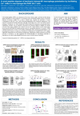
CSP32 activated the MAPKs and NF-κB CSP32 stimulated cytosolic Ca 2+ influx CSP32-mediated Ca 2+ influx regulated M1
signaling pathways in macrophages during M1 polarization polarization
Figure 4. CSP32 activated MAPKs and the NF-κB signaling pathway in Figure 5. CSP32 upregulated cytosolic Ca2+ concentrations during M1 polarization. Figure 6. Ca 2+ regulated CSP32-mediated M1 polarization. (A-C) Cells were
macrophages. (A) At 0, 15 min, 30 min, 1 h, 2 h and 24 h after CSP32 treatment, Cells were treated with the indicated concentrations of CSP32 and LPS for 24 h. pretreated with or without 5 μM BAPTA-AM for 30 min and then incubated with the
cells were harvested and lysed. (B, C) Cells were treated with 1 μg/mL and 5 μg/mL Then, cells were stained with 2 μM fluo-3-AM for 30 min. (A) Representative indicated concentrations of CSP32 and LPS for 24 h. (A) The amount of NO in the
CSP32 for 1 h and subsequently harvested and lysed. (A, B) Total cell lysates were histogram from the flow cytometric analysis of fluo-3A-M-positive macrophages cell supernatants was measured using Griess reagents. Data are expressed as the
examined by Western blotting for ERK, JNK and p38 MAPK phosphorylation. β- following CSP32 and LPS treatment. (B) Representative fluorescence images. mean ± SD (n=4). *p <0.05 and **p <0.01 compared between the groups. (B)
actin was used as an internal control. (C) Cytoplasmic and nuclear lysates were Scale bar; 20 μm. (C) At 0, 15 min, 30 min, 1 h, 2 h and 24 h after CSP32 treatment, Representative flow cytometric histogram of the phagocytosis capacity using
examined by Western blotting for IκB-α and NF-κB. Actin and lamin B1 serve as the cells were harvested and lysed. Total cell lysates were examined by Western fluorescent FITC-IgG latex beads. (C) Protein expression of iNOS. β-actin was used
internal controls for the cytoplasmic and nuclear lysates, respectively. (D) In nuclear blotting for PLCγ1, PKCα, PKCβ and PKCε. β-actin was used as an internal control. as an internal control for total cell lysates. (D, E) Cells were pretreated with or
lysates, NF-κB activity was analyzed using an NF-κB p65 transcription factor assay without 5 μM BAPTA-AM for 30 min and then incubated with the indicated
kit. Data are expressed as the mean ± SD (n=4). *p <0.05 and **p <0.01 compared concentration of 5 μg/mL CSP32 for 1 h. (D) Cytoplasmic and nuclear lysates were
with the control. examined by Western blotting for IκB-α and NF-κB. Actin and lamin B1 serve as the
internal controls for the cytoplasmic and nuclear lysates, respectively. (E)
Localization of NF-κB p65 (green) was visualized with fluorescence microscopy.
Cells were stained with DAPI for visualization of the nuclei (blue).
METHODS CONCLUSION
REFERENCES
Cell viability: MTT assay
Measurement of nitric oxide (NO) : Griess reagent 1. Parihar, A.; Eubank, T.D.; Doseff, A.I. Monocytes and macrophages regulate
immunity through dynamic networks of survival and cell death. J. Innate. Immun.
Phagocytosis analysis: phagocytosis assay 2010, 2, 204‐215.3
2. Wu, X.Q.; Dai, Y.; Yang, Y.; Huang, C.; Meng, X.M.; Wu, B.M.; Li, J. Emerging
Cytokine expression: Gene expression microarray analysis, role of microRNAs in regulating macrophage activation and polarization in
immune response and inflammation. Immunology 2016, 148, 237‐248.
ELISA kits and WB 3. Choi, Y.H.; Cho, S.S.; Simkhada, J.R.; Rahman, M.S.; Choi, Y.S.; Kim, C.S.;
Yoo, J. C. A novel multifunctional peptide oligomer of bacitracin with possible
Inflammatory response : RT-PCR, WB, Nuclear fraction bioindustrial and therapeutic applications from a Korean food-source Bacillus
strain. PLoS One 2017, 12, e017697118. 18.
and IF 4. Kwon, D.H.; Lee, H.; Park, C.; Hong, S.H.; Hong, S. H.; Kim, G.Y.; Cha, H.J.;
Kim, S.; Kim, H.S.; Hwang, H. J.; Choi, Y.H. Glutathione induced immune-
Intracellular calcium analysis: Fluo-3-AM assay CSP32 stimulates M1 polarization via the calcium-dependent NF-κB signaling pathway stimulatory activity by promoting M1-like macrophages polarization via potential
ROS scavenging capacity. Antioxidants (Basel) 2019, 8, 413.