
Page 15 - F. Cell biology
P. 15
Identification of small molecules that can inhibit necroptotic cell
death
Su Bin Sin¹ and Young Sik Cho¹*
College of Pharmacy, Keimyung University, 1000 Sindang-dong, Dalseo-gu, Daegu 704-701, Republic of Korea
BACKGROUND AIM
Necroptosis or programmed necrosis is a specialized and regulated necrosis. Programmed necrosis (necroptosis) has been recognized as a specialized and
regulated necrosis that is aroused when apoptosis is defective. Initially, it is
It has been proposed that necroptotic cell death is pathologically associated known to play a beneficial significance in innate immune response to viral
with ischemic brain injury and retinal disorder. infection that can evade immune surveillance. Now, it has been emerging as
the strategy to overcome the cancers with acquired drug resistance. In
Necrostatin-1 (Nec-1) was for the first time identified as an inhibitor of contrast, it has been proposed that necroptotic cell death is pathologically
receptor interacting protein 1 (RIP1), a necroptosis regulator. associated with ischemic brain damage and degenerative diseases. Here, as
an effort to discover hits that can selectively inhibit necroptotic cell death,
With growing knowledge of necroptosis-associated diseases, needs for we screened in-house and in silico chemical libraries in a cell based assay.
therapeutic drugs are being pursued. Eventually, 7 hits were identified from in-house chemical library while 2
hits were from modeling data. Mostly, hits exhibited less protective activity
To discover hits that could rescue cells from necroptotic strimuli, we set out to from TNF- and zVAD-mediated necroptosis than a reference compound
screen in-house chemical library (6800 chemicals) and computer modeling- Nec-1. Interestingly, a few of hits had preferential protective effects on
predicted compounds in the necroptosis-prone cells L929. zVAD or TNFα while Nec-1 exhibited similar IC50 against zVAD or TNF,
suggesting that deduced chemicals can discriminate signaling pathways
leading to receptor or nonreceptor-mediated necroptotic cell death.
RESULTS
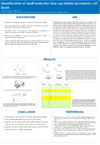
Figure 4. Summary of screening outcomes for discovery of hits with antinecroptotic potency.
The dotted line represents protection cutoff (50%) drawn for the primary screen of hit
candidates. Compounds suggested from a chemical library or computer modeling were assayed
for protective activity against TNFα-mediated necroptosis in L929 cells. Each compound from
chemical sources was sorted in the decreasing order of protective potency (A). Probability of
selecting compounds with protective activity of more than 50% are approximately 0.12% and
Figure 1. Schematic flow chart for isolation of hits with antinecroptotic activity selectively. To find hits that 1.28%, respectively, from a chemical library and computer modeling-deduced chemicals. When
can specifically protect cells from necroptotic stress, L929 cells, which were pretreated with various compounds compounds were sorted by protective percentage (B), a few of compounds effectively
out of a chemical libraryfor 1 h, were stimulated with TNFα to induce necroptotic cell death. protected cells from TNFα-mediated necroptosis, representing that only 15 and 2 compounds,
respectively, exerted protective activity of more than 50% out of chemicals from different
sources.
Figure 2. The binding mode of compound 9 against RIP1.The green lines represents H-bond.
Table 1. Antinecroptotic activities of hits selected from screening of in-
house chemical library and computer modeling-deduced chemicals. In-
house chemical library was from KRICT, including about 6800 chemicals
classified by representative chemical moiety. Also, some chemicals were
Figure 3. Cell-based assays suitable for apoptosis and programmed necrosis (necroptosis). Induction suggested from database of chembank (KRICT) through computer modeling.
and protection of cell death were tested in 3 different cell lines including L929, HeLa and NIH3T3 under the Nine compounds selected from primary screening were further assayed to
various conditions of cell death stimuli (A). Expression levels of RIP1 and RIP3, key necroptotic regulators, plot dose-response curves against TNFα- or zVAD-driven necroptosis, and
were investigated in L929, HeLa and NIH3T3 (B) then EC 50 of each compound was calculated using GraphPad Prism software.
CONCLUSION REFERENCES
1. Establishment of a cell based assay for screening antinecroptotic chemicals 1. Han W, Li L, Qiu S, Lu Q, Pan Q, Gu Y, Luo J, Hu X. Shikonin circumvents cancer
drug resistance by induction of a necroptotic death. Mol Cancer Ther 2007; 6: 1641-
2. 9 hits were finally discovered and those structures were released 1649.
3. Some hits protected cells from both zVAD and TNFα-mediated necroptosis, 2. Park SY, Shim JH, Cho YS. Distinctive roles of receptor-interacting protein kinases 1
and 3 in caspase-independent cell death of L929. Cell Biochem Funct 2014; 32: 62-69.
while a few hits had preferential protective effects on zVAD or TNF α 3. Cho YS, Challa S, Moquin D, Genga R, Ray TD, Guildford M, Chan FK.
Phosphorylation-driven assembly of the RIP1-RIP3 complex regulates programmed
4. Especially, Compound 9, which exhibited high similarity score (FCFC4>0.7), necrosis and virus-induced inflammation. Cell 2009; 137: 1112-1123.
is comparable to Nec-1, a RIP1 specific inhibitor 4. He S, Wang L, Miao L, Wang T, Du F, Zhao L, Wang X. Receptor interacting protein
kinase-3 determines cellular necrotic response to TNF-alpha. Cell 2009; 137: 1100-
1111.
5. Zhang DW, Shao J, Lin J, Zhang N, Lu BJ, Lin SC, Dong MQ, Han J. RIP3, an energy
metabolism regulator that switches TNF-induced cell death from apoptosis to necrosis.
Science 2009; 325: 332-336.