
Page 11 - F. Cell biology
P. 11
Anticancer effect of an IRAK4 inhibitor on ABC-DLBCL cells
Su Bin Sin¹ and Young Sik Cho¹*
College of Pharmacy, Keimyung University, 1000 Sindang-dong, Dalseo-gu, Daegu 704-701, Republic of Korea
BACKGROUND AIM
TLR/IL-1R Potent interleukin-1 receptor-associated kinase 4 (IRAK4) inhibitors have been
emerging as a blockade of toll-like receptor/ I interleukin-l receptor (TLR/ILR)
MYD88 signaling pathway to treat autoimmune-related diseases such as rheumatoid
Myddosome
IRAK4 arthritis and multiple sclerosis. Additionally, some cancers have been reported to
show an increase in IRAK4 activity or to bear L265P mutation in MyD88, an
P IRAK1 P intracellular adaptor protein of TLR/ILlR, resulting in constitutive activation of a
Complex Ⅰ TAK1
TRAF6 cascade of downstream signaling pathway. A hit compound 6-(imidazo 1, 2-a]
pyridine-3-yl)-N-(4-piperidinyl)-2-pyridinamine was initially discovered as a JNK
IKK
kinase inhibitor, but was later found to have considerable potency in an in vitro
enzymatic assay with IC50 of 216 nM. To delineate the underlying mechanism of
IkB
p65 p50 IRAK4 target-based inhibition of lymphoma proliferation, this compound was further
evaluated in cell-based assays. An IRAK4 inhibitor potently down-regulated not only
p65 p50 the LPS-induced NFκB transcriptional activity in NFκB-luciferase A549 cell line, but
also NO production in LPS-stimulated RAW264.7.
Toll-like receptor (TLR) is one of pathogen recognition receptors(PRRs) leading to innate immune
responses
Furthermore, a subgroup of DLBCL OCI-Ly3 cell line bears a mutation in MyD88 so
IRAK4 is a crucial protein for expression of pro-inflammatory cytokine and cell survival proteins via that it can aggressively proliferate in clumps even though not being stimulated.
activation of NF-κB Treatment of OCI with IRAK4 inhibitor reduced cell viability in a dose dependent
IRAK4 is a down stream protein of an adaptor protein MyD88 in TLR signaling pathway. manner, and induced an increase in cell population undergoing early apoptosis and
late apoptosis with treatment times. Therefore, IRAK4 targeting is proposed to be a
IRAK4 hyperphosphorylates IRAK1 and then activates the downstream signaling of TLR, consequently promising strategy in inhibiting the proliferation of MyD88-defective lymphoma in
activating NF-κB and AP-1 transcription factors. addition to treatment of autoimmune-related diseases.
6-(imidazo[1,2-a]pyridine-3-yl)-N-(4-piperidinyl)-2-pyridinamine (CIM741) is a hit compound targeting
IRAK4
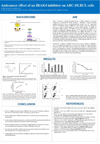
RESULTS
4 120%
(A) Structure of CIM741 (B) IC 50 o f #741 in en z ym e assa y 100%
100 3
N 741 80%
D M SO 71% inhibition
N H N 75 Absorbant (fold of control) 2 60% ***
N 50 1 Cell viability (% of control) 40% ***
N 25 20%
H 0
0 - - #741 #800 #802 #803 0%
5
1
-7 .5 -7 .0 -6 .5 -6 .0 -5 .5 -5 .0 -4 .5 LPS (+) 0 Concentration (uM) 10
logM Figure 3. Inhibition of NO production by IRAK4 inhibitor. RAW246.7 cells plated in 96-
Figure 1. In vitro enzymatic assay of IRAK4 inhibitor CIM741. well plate were pre-treated with different chemicals at 10μM for 30min and then Figure 4. Dose-dependent inhibition of cell proliferation by CIM741. OCI-Ly3 cells were
(A) Structure of CIM 741 stimulated with LPS (1 μg/ml) for 18h. Supernatants (100 μl) taken from each well were seeded at a density of 2×10 4 onto each well of 96-well plate and then treated with
compounds from 1 to 10 μM for 48 h. Proliferation was measured by staining cells with
transferred to another 96-well plate. After Griess reagent (100 μl) mixed with the
(B) A dose-response curve for IRAK4 inhibition were plotted with increasing doses of 741. transferred supernatants, the absorbance of the reaction mixture was measured at WST-1. Error bars are the mean ± S.D. This was performed in triplicate. (*p < 0.05, **p
IRAK4 enzyme was incubated with increasing concentrations of 741 compound, ranging 540nm. < 0.01 and ***p < 0.001 vs. 0 μM CIM741 treated group).
from 0.5 to 10 μM for 30min at 30℃.
% inhibition in luciferase assay
120 NS #741
DMSO DMSO NS stain - All Events 741 - All Events
100 CIM741 10 5 UL UR 5 UL UR
#741
19.08 15 … 10 24.71 40.91
80
% inhibiton 60 Propidium Iodide-A 10 10 4 3 Propidium Iodide-A 10 10 4 3
20 40 10 2 0 LL LR 10 0 2 LL LR NS
64.40 1.52 30.33 4.05
0 -10 2 0 10 2 only Z - All Events 10 4 10 5 0 10 2 741+Z - All Events FITC-A 10 4 10 5
10 3
10 3
FITC-A
-7.0 -6.5 -6.0 -5.5 -5.0 -4.5 PI 5 UL UR 5 UL UR
concentration (logM) 10 24.58 18.32 10 21.57 29.96
Figure 2. IC 50 determination of CIM741 for suppression of NFκB activity in A549/NFκB- 10 4 10 4
luciferase cells. Cells were plated at a density of 1×10 4 to each well of 96-well plate. A549 Propidium Iodide-A 10 3 Propidium Iodide-A 10 3
bearing NFκB-luciferase reporter gene were pre-treated with CIM741 from 0.1 to 10 μM 2 10 2 zVAD Figure 5. CIM741-mediated cell death mode in OCI-Ly3 cells. Cells
for 30 min and then stimulated with LPS (1 ㎍/1 ㎖) for 4 h. Cells were rinsed with PBS, 10 0 LL 56.01 LR 1.09 0 LL 45.38 LR 3.09 were plated at a density of 2x10 5 onto each well of 12well and pre-trated with
lysed by lysis buffer (125 mM Tris-H 3 PO 4 (pH 7.8), 10 mM EDTA, 10 mM DTT, 50% glycerol, -10 2 0 10 2 10 3 FITC-A 10 4 10 5 -10 2 0 10 2 10 3 FITC-A 10 4 10 5 zVAD (10μM) and then vehicle or a IRAK4 inhibitor CIM741 (10μM) for 48h.
and 5% TritonX-100) for 5 min and detected by using luciferase substrate. Then, cells were stained with annexinV and PI to do flow cytometricanalysis.
FITC
CONCLUSION REFERENCES
1. Beutler, B., et al. (2004). “Innate immunity: an overview.” Mol Immunol 40(12):
845-859.
CIM741 inhibited enzymatic activity of IRAK4 in vitro assay which measured indirectly 2. Janeway, C. A., Jr., et al. (2002). “Innate immune recognition.” Annu Rev Immunol
ADP used up for the kinase reaction, showing IC 50 value of 216 nM. 20: 197-216.
3. Rock, F. L., et al. (1998). “A family of human receptors structurally related to
CIM741 significantly inhibited NF-κB activation in luciferase assay by interfering with Drosophila Toll.” Proc Natl Acad Sci USA 95(2): 588-593.
IRAK4 kinase. 4. Medzhitov, R., et al. (1997). :A human homologue of the Drosophila Toll protein
signals activation of adaptive immunity.” Nature 388(6640): 394-397.
NF-κB activation and iNOs production in cells treated CIM741 was decreased to 80% and 5. Fekonja, O., et al. (2012). "Suppression of TLR signaling by targeting TIR domain-
71%, respectively, when compared with vehicle-treated group. containing proteins." Curr Protein Pept Sci 13(8): 776-788.
6. Chaudhary, D., et al. (2015). "Recent advances in the discovery of small molecule
CIM741-mediated cell death was induced through activation of caspase 3/7, and was inhibitors of interleukin-1 receptor-associated kinase 4 (IRAK4) as a therapeutic
partially protected by a pancaspase inhibitor zVAD (data not shown). target for inflammation and oncology disorders." J Med Chem 58(1): 96-110.
7. Ferreri, A. J., et al. (2009). "Infectious agents and lymphoma development:
From the flow cytometric analysis, there was an increase of dead cell population when molecular and clinical aspects." J Intern Med 265(4): 421-438.
treated with CIM741, as compared with control. 8. Rhyasen, G. W. and D. T. Starczynowski (2015). "IRAK signalling in cancer." Br J
Cancer 112(2): 232-237.
Targeting IRAK4 may be an effective strategy for inhibition of proliferative lymphoma
with MyD88 mutant.