
Page 9 - F. Cell biology
P. 9
Identification of ANO4/TMEM16D functions in pterygium
1
Jiyeon Kim , Ikhyun Jun , and Seo Kyoung Yul 1,2
2
1 Brain Korea 21 PLUS for Medical Sciences, Yonsei University, Seoul, Korea
2 Department of Ophthalmology, Yonsei University College of Medicine, Seoul, Korea
BACKGROUND AIM
The Anoctamin (ANO)/transmembrane member 16 (TMEM16) family genes mediate diverse
physiological and pathophysiological functions including cancer cell proliferation. In particular, they The ANO4 appears to be a clinically useful
induce cancer cell growth through interaction with epidermal growth factor receptor (EGFR). prognostic marker for pterygium and a potential
therapeutic target. Surgical excision is a standard
In this study, we investigate the role of ANO4/TMEM16D in proliferation of pterygium. Pterygium is an
ocular surface disease and fibrovascular tissue of the conjunctiva grows excessively like cancer, treatment for pterygium and possibility of
covering the cornea. In an initial screen of ANO/TMEM16s, ANO4/TMEM16D was overexpressed in recurrence after surgical removal has been
reported to range from 38% to 88% according to
pterygium and its role was evaluated using in vitro approach. We observed overexpression of ANO4 in
pterygium tissue compared to normal conjunctiva tissue. In addition, physical association of ANO4 the study. It can be also developed as a
medication to prevent recurrence after surgery.
with EGFR underlies ANO4-induced cell proliferation, like ANO1, ANO9 by mechanical analysis.
METHODS
1. Quantitative PCR analysis. Quantitative PCR (qPCR) was performed using the Applied Biosystems StepOne system (Applied Biosystems). According to the comparative
threshold cycle ( ) method (Livak and Schmittgen, 2001), expression of the target gene was normalized to the expression of the housekeeping gene glyceraldehyde 3-
phosphate dehydrogenase (GAPDH), yielding the delta (d ) value.
2. Tissue Immunofluorescence. Frozen blocks containing conjunctiva, pterygium tissues were sectioned at 6 μm thickness and sections were blocked with 5% donkey serum
and 1% BSA in 1X PBS containing 0.1% Triton X-100 (Sigma) for 1 h, and then incubated with primary antibodies against ANO4 diluted in the blocking solution at 4℃
overnight. After washing with 1X PBS, the sections were incubated with Alexa Fluor 488-conjugated secondary antibodies at room temperature 1 h. After washing, slides
were then mounted with Vectashield Mounting Medium (VECTOR LABORATORIES).
3. WST-8 Assay. For cell viability assay, conjunctival epithelial cells, fibroblasts were transfected using Lipofectamin LTX with PLUS reagent (Invitrogen). 72 h post transfection,
cell proliferation was determined by water-soluble tetrazolium salt-8 (WST-8) assay using Quanti-MAX WST-8 cell viability assay kit (Biomax Inc), according to the
™
4
instructions. Conjunctival epithelial cells, fibroblasts were plated at 2 × 10 cells/ml in 96-well plate. Subsequently, 10 μL of WST-8 solution was added to each well, and
incubated 2 h. Absorbance at 450 nm was measured by microplate reader.
4. Immunoprecipitation. For the immunoprecipitation assay, PANC-1 cells were co-transfected with the ANO1-myc, ANO5-myc, ANO6-myc, ANO9-myc, and EGFR-HA plasmids.
The cell lysates were mixed with anti-myc antibodies and incubated overnight at 4 °C in lysis buffer. The immune complexes were collected by binding to protein G/A-
Sepharose and washed three times with lysis buffer. The immunoprecipitates and lysates were then separated by SDS–PAGE and immunoblotted with anti-HA antibodies.
RESULTS
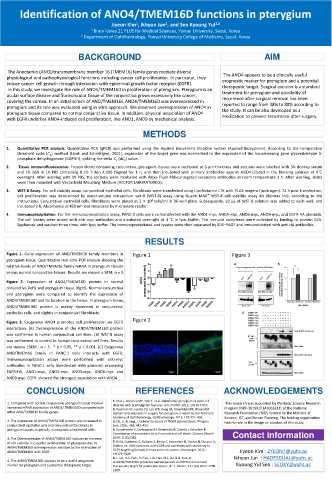
Figure 1. Gene expression of ANO/TMEM16 family members in Figure 1 Figure 3
pterygium tissue. Quantitative real-time PCR analysis showing the
relative levels of ANO/TMEM16 family mRNA in pterygium tissues
versus normal conjunctiva tissues. Results are means ± SEM; n = 5.
Figure 2. Expression of ANO4/TMEM16D protein in normal
conjunctiva (left) and pterygium tissue (right). Normal conjunctiva
and pterygium were compared to identify the expression of
ANO4/TMEM16D and its location in the tissue. In pterygium tissue,
ANO4/TMEM16D protein is mainly expressed in conjunctival
epithelial cells, and slightly in conjunctival fibroblasts. C
Figure 2
Figure 3. Exogenous ANO4 promotes cell proliferation via EGFR
interaction. (A) Overexpression of the ANO4/TMEM16D protein
was confirmed in human conjunctival cell lines. (B) WST-8 assay
was performed to control in human conjunctival cell lines. Results
are means ±SEM ; n = 3. * p < 0.05, ** p < 0.001. (C) Exogenous
ANO/TMEM16 family in PANC-1 cells interacts with EGFR.
Immunoprecipitation assays were performed with anti-myc
antibodies in PANC-1 cells transfected with plasmids expressing
EGFR-HA, ANO1-myc, ANO4-myc, ANO5-myc, ANO6-myc and
ANO9-myc. EGFR showed the strongest association with ANO4.
CONCLUSION REFERENCES ACKNOWLEDGEMENTS
1. Chui J, Coroneo MT, Tat LT, et al. Ophthalmic pterygium: A stem cell
1. Compared with normal conjunctiva, pterygium tissue showed disorder with premalignant features. Am J Pathol. 2011; 178:817-827. This research was supported by the Basic Science Research
increased mRNA expression of ANO4/ TMEM16D compared with 2. Kaufman SC, Jacobs DS, Lee WB, Deng SX, Rosenblatt MI, Shtein RM. Program (NRF-2019R1F1A1063311) of the National
other ANO/TMEM16 family genes. Options and adjuvants in surgery for pterygium: a report by the American Research Foundation (NRF) funded by the Ministry of
Academy of Ophthalmology. Ophthalmology. 2013; 120:201–208. Science, ICT, and Future Planning. The funding organization
2. The expression of ANO4/TMEM16D protein was increased in 3. Oh, U. & Jung, J. Cellular functions of TMEM16/anoctamin. Pflügers had no role in the design or conduct of this study.
conjunctival epithelial cells and conjunctival fibroblasts in Arch.2016; 468, 443–453.
pterygium tissues, especially in conjunctival epithelial cells. 4. Kunzelmann K, Ousingsawat J, Benedetto R, Cabrita I, Schreiber R.
Contribution of anoctamins to cell survival and cell death. Cancers (Basel). Contact information
3. The Overexpression of ANO4/TMEM16D induces an increase 2019; 11(3):E382.
of cell viability. It can infer proliferation of pterygium due to 5. Bill A, Gutierrez A, Kulkarni S, Kemp C, Bonenfant D, Voshol H, Duvvuri U,
ANO4/TMEM16D overexpression and due to the interaction of Gaither LA. ANO1 interacts with EGFR and correlates with sensitivity to
ANO4/TMEM16D with EGFR. EGFR-targeting therapy in head and neck cancer. Oncotarget. 2015; Jiyeon Kim : ZYEON7@yuhs.ac
6:9173–9188.
6. I. Jun, H.S. Park, H. Piao, J.W. Han, M.J. An, B.G. Yun, et Ikhyun Jun : HADESDUAL@yuhs.ac
4. The ANO4/TMEM16D appears to be a useful prognostic al.ANO9/TMEM16J promotes tumourigenesis via EGFR and is a novel
marker for pterygium and a potential therapeutic target. therapeutic target for pancreatic cancer, Br. J. Cancer, 117 (12) 2017; 1798- Kyoung Yul Seo : SEOKY@yuhs.ac
1809.