
Page 5 - F. Cell biology
P. 5
Novel degradation mechanism of c-Fos by
AKT2-mediated autophagy pathway
1
1
1
1,#
1,#
Wooram Choi , Deok Jeong , Han Gyung Kim , Yo Han Hong , Chaoran Song , Long You , Jieun Oh , and Jae Youl Cho 1
1
1
1 Department of Integrative Biotechnology, Sungkyunkwan University, Suwon 16419, Korea
BACKGROUND AIM
Autophagy is an important autolysis process that responds to a
variety of stresses and maintains intracellular balance, contributing to To figure out the role of AKT2 in c-Fos degradation thr
cell homeostasis. Although AKT is an important signaling protein in ough autophagy pathway.
the cell pathway, its role is not fully understood in autophagy.
METHODS
Expression levels of c-Fos, c-Jun, LC3B, ATG12, and AKT2 in RAW264.7 cell and HEK293T cell were evaluated by western blot.
And luciferase assay was used to assess the transcriptional activity of c-Fos in various experimental conditions. And to
confirm the interaction of c-Fos and AKT2, HEK293T cell overexpressing both of AKT2 and c-Fos were made and the samples
were evaluated by co-immunoprecipitation.
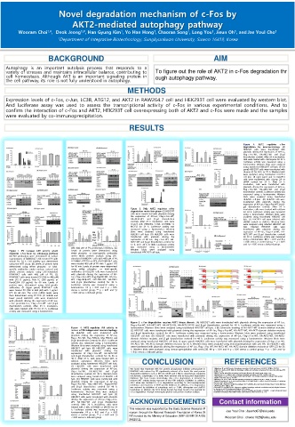
RESULTS
Figure 4. AKT2 regulates c-Fos
degradation by macroautophagy (A)
HEK293T cells were transfected with
plasmids driving the expression of AP-Luc,
Flag-c-Fos-WT, HA-AKT2-WT and β-gal
(transfection control). After 24 h incubation,
cells were treated with chloroquine for 24 h.
Luciferase activity was measured using a
luminometer. Western blots were analyzed
using transfected HEK293T cell lysis. (B left
panel) HEK293T cells were transfected with
siLamp 2A for 48 h or 72 h. Western blots
were analyzed using transfected HEK293T
cell lysis. (B right panel and C) HEK293T
cells were transfected with siLamp 2A or
siRNA control for 24 h. After 24 h
incubation, cells were transfected with
plasmids driving the expression of AP-Luc,
Flag-c-Fos-WT, HA-AKT2-WT and β-gal
(transfection control). Luciferase activity was
measured using a luminometer. Western
blots were analyzed using transfected
HEK293T cell lysis. (D) HEK293T cells were
transfected with plasmids driving the
expression of AP-Luc, Flag-c-Fos-WT and β-
gal (transfection control). After 24 h
Figure 3. Only AKT2 regulates c-Fos incubation, cells were treated with rottlerin
degradation under late phase (C) HEK293T for 24 h. Luciferase activity was measured
cells were transfected with plasmids driving using a luminometer. Western blots were
the expression of AP-Luc, Flag-c-Fos-WT, analyzed using transfected HEK293T cell
HA-AKT2-WT and β-gal (transfection lysis. (E) HEK293T cell were infected with
control). After 24 h incubation, cells were shRNA control or shATG5. Western blots
treated with BN82002, 3-MA, LY294002 or were analyzed using infected HEK293T cell
MG132 for 24 h. Luciferase activity was lysis. Infected HEK293T cells were
measured using a luminometer. Western transfected with plasmids driving the
Figure 2. PV increase AKT activity which blots were analyzed using transfected expression of AP-Luc, Flag-c-Fos-WT, HA-
degrades c-Fos level by autophagy pathway HEK293T cell lysis. (D) HEK293T cells were AKT2-WT and β-gal (transfection control)
(Continued) (E) Levels of protein were transfected with plasmids driving the for 48 h. Luciferase activity was measured
determined using either phospho- or total- expression of AP-Luc, Flag-c-Fos-WT, HA- using a luminometer. ## p < 0.01 and # p
specific antibodies under whole protein AKT2-WT and β-gal (transfection control for < 0.05 versus a control group, ** p < 0.01
analysis using LPS-stimulated RAW264.7 cells 12 h, 24 h ,36 h or 48h). Luciferase activity and *p < 0.05 versus a induced group.
with 400 µM of PV, proteolysis inhibitors. (G) was measured using a luminometer.
Figure 1. PV increase AKT activity which Levels of protein were determined using Western blots were analyzed using
transfected HEK293T cell lysis.
degrades c-Fos level by autophagy pathway either phospho- or total-specific antibodies
(A) NO production were determined in culture under whole protein analysis using LPS-
supernatants of RAW264.7 cells treated PV with stimulated RAW264.7 cells with 400 µM of PV,
stimuli for 24 h. Cell viability was determined LY294002, KT5720 or GFX. (H) RAW264.7 cells
using the MTT assay. (B) Levels of protein were were treated 400 µM of PV with 1 µg/mL LPS
determined using either phospho- or total- for 60 min. Levels of protein were determined
specific antibodies under nuclear, cytosol and using either phospho- or total-specific
whole protein analysis using LPS-stimulated antibodies. (I) HEK293T cells were transfected
RAW264.7 cells with 400 µM of PV. (C) with plasmids driving the expression of AP-
RAW264.7 cells were treated PV (200 to 400 µM) Luc, Flag-c-Fos-WT, Flag-Myd88-WT, HA-Src-
with LPS (1 µg/mL), Poly I:C (200 µg/mL) or WT, Flag-IKKβ-WT, CFP-TRIF-WT, HA-TBK1
Pam3CSK4 (10 µg/mL) for 60 min. Levels of and β-gal (transfection control) for 48 h.
protein were determined using total-specific Luciferase activity was measured using a
antibodies. (D Upper panel) RAW264.7 cells luminometer. ## p < 0.01 and # p < 0.05
were treated PV (200 to 400 µM) with 1 µg/mL versus a control group, ** p < 0.01 and *p
LPS for 60 min. The c-Fos mRNA levels were < 0.05 versus a induced group.
then determined using RT-PCR. (D middle and
lower panel) HEK293T cells were transfected
with plasmids driving the expression of AP-Luc,
c-Fos promoter-Luc, Flag-c-Fos-WT and β-gal
(transfection control). After 24 h incubation,
cells were treated with PV for 24 h. Luciferase
activity was measured using a luminometer.
Figure 5. c-Fos degradation requires AKT2 kinase domain. (A) HEK293T cells were transfected with plasmids driving the expression of AP-Luc,
Flag-c-Fos-WT, HA-AKT2-WT, HA-AKT2-DN, HA-AKT2-S474D and β-gal (transfection control) for 48 h. Luciferase activity was measured using a
Figure 6. AKT2 regulates JNK activity to luminometer. Western blots were analyzed using transfected HEK293T cell lysis. ) (B) Schematic drawing of HA-AKT2-WT domain deletion mutants.
induce mTOR independent macroautophagy. (C) HEK293T cells were transfected with plasmids driving the expression of AP-Luc, Flag-c-Fos-WT, HA-AKT2-WT, HA-AKT2-domain deletion mutants
(A) HEK293T cells were transfected with and β-gal (transfection control) for 48 h. Luciferase activity was measured using a luminometer. Western blots were analyzed using transfected
plasmids driving the expression of AP-Luc, HEK293T cell lysis. (D) HEK293T cells were transfected with plasmids driving the expression of AP-Luc, Flag-c-Fos-WT, HA-AKT2-WT, HA-AKT2-
Flag-c-Fos-WT, Flag-PI3KCA, Myc-PI3KC3 and domain deletion mutants and β-gal (transfection control) for 48 h. Luciferase activity was measured using a luminometer. Western blots were
β-gal (transfection control) for 48 h. Luciferase analyzed using transfected HEK293T cell lysis. (E upper panel) HEK293T cells were transfected with plasmids driving the expression of Flag-c-Fos-WT,
activity was measured using a luminometer. HA-AKT2-WT, HA-AKT2-domain deletion mutants for 36 h. Western blots were analyzed using immunoprecipitation with anti-HA.. (F) HEK293T cells
Western blots were analyzed using transfected were transfected with plasmids driving the expression of AP-Luc, Flag-c-Fos-WT, HA-AKT2-WT, HA-AKT2-domain deletion mutants or GFP-LC3 for 36
HEK293T cell lysis (B) HEK293T cells were h. Western blots were analyzed using immunoprecipitation with anti-LC3B. ## p < 0.01 and # p < 0.05 versus a control group, ** p < 0.01 and *p
transfected with plasmids driving the
expression of Flag-c-Fos-WT, HA-AKT2-WT < 0.05 versus a induced group.
and β-gal (transfection control) for 24, 36 or
48 h. After 24 h, cells were treated with or
without rapamycin. Western blots were CONCLUSION REFERENCES
analyzed using transfected HEK293T cell lysis.
(C) HEK293T cells were transfected with
plasmids driving the expression of AP-Luc, We found that treatment with the protein phosphatase inhibitor pervanadate in 1.Mizushima, N. and M. Komatsu, Autophagy: renovation of cells and tissues. Cell, 2011. 147(4): p. 728-741.
Flag-c-Fos-WT, HA-AKT2-WT and β-gal RAW264.7 cells induced by LPS significantly reduced c-Fos levels. We used protein 2.Glick, D., S. Barth, and K.F. Macleod, Autophagy: cellular and molecular mechanisms. The Journal of pathology, 2010.
221(1): p. 3-12.
(transfection control) for 48 h. Western blots degradation inhibitors such as MG132, AICAR and 3-MA to demonstrate regulatory 3.Jiang, P. and N. Mizushima, Autophagy and human diseases. Cell research, 2014. 24(1): p. 69.
4.Rubinsztein, D.C., G. Mariño, and G. Kroemer, Autophagy and aging. Cell, 2011. 146(5): p. 682-695.
were analyzed using transfected HEK293T cell mechanisms. Surprisingly, c-Fos levels were restored only by lysosomal degradation 5.Halazonetis, T.D., et al., c-Jun dimerizes with itself and with c-Fos, forming complexes of different DNA binding
lysis. (D) HEK293T cells were transfected with inhibitors. We have found, interestingly, that c-Fos degradation is restored by affinities. Cell, 1988. 55(5): p. 917-924.
6.Manning, B.D. and A. Toker, AKT/PKB signaling: navigating the network. Cell, 2017. 169(3): p. 381-405.
plasmids driving the expression of AP-Luc, reduced AKT activity during exposure of the PI3K inhibitor LY294002. In addition, 7.Matsuda-Lennikov, M., et al., Lysosomal interaction of Akt with Phafin2: a critical step in the induction of autophagy.
AKT2 alone was involved in c-Fos degradation according to AKT2-overexpression
PloS one, 2014. 9(1): p. e79795.
Flag-c-Fos-WT, Myc-JNK2-WT, Flag-p38-WT conditions and treatment of AKT2-specific inhibitors. In addition, c-Fos degradation 8.Cuervo, A.M., Chaperone-mediated autophagy: Dice's' wild'idea about lysosomal selectivity. Nature reviews Molecular
and β-gal (transfection control) for 48 h. was restored by inhibition of autophagy such as 3-MA, chloroquine and shATG5 cell biology, 2011. 12(8): p. 535.
9.Orenstein, S.J. and A.M. Cuervo. Chaperone-mediated autophagy: molecular mechanisms and physiological relevance.
Luciferase activity was measured using a knockdown. In addition, the AKT2 kinase domain has been found to be essential for in Seminars in cell & developmental biology. 2010. Elsevier.
10. Hanada, M., J. Feng, and B.A. Hemmings, Structure, regulation and function of PKB/AKT—a major therapeutic target.
luminometer. Western blots were analyzed c-Fos degradation through immunoprecipitation and protein mutation analysis. Biochimica et Biophysica Acta (BBA)-Proteins and Proteomics, 2004. 1697(1-2): p. 3-16.
using transfected HEK293T cell lysis (E)
HEK293T cells were transfected with plasmids
driving the expression of AP-Luc, Flag-c-Fos- ACKNOWLEDGEMENTS Contact information
WT, HA-JNK2-WT and β-gal (transfection
control). After 24 h incubation, cells were
treated with 3-MA, LY294002 or MG132 for 24 This research was supported by the Basic Science Research P
h. Luciferase activity was measured using a Jae Youl Cho : Jaecho67@skku.edu
luminometer. ## p < 0.01 and # p < 0.05 rogram through the National Research Foundation of Korea (N
versus a control group, ** p < 0.01 and *p RF) funded by the Ministry of Education (NRF-2016R1D1A1B0
< 0.05 versus a induced group. Wooram Choi : chwoo1028@skku.edu
3932512).