
Page 87 - D. Cancer biology
P. 87
[D-58] Significance of Jagged-1 activated by APEX1 as chemoresistance
factors in gastric cancer
Hong-Beum Kim
Department of Premedical Course, Chosun University School of Medicine, 309 Pilmun-daero, Dong-gu,
Gwang-ju 61452,Republic of Korea
Abstract Background/Aim: We investigated the clinical role of the molecular targets, APEX1 and Jagged-1, and the Apex1 - Jagged-1 cascade in
gastric cancer cells. Materials and Methods: We used 6 human gastric cancer cell lines (SNU-1, SNU-5, SNU-16, NCI-N87, KATO- III and
AGS), and demonstrated the chemosensitivity of APEX1 and Jagged-1 through the MTT assay and immunoblotting. Tumor growth was
assayed following cisplatin and 5-FU treatment using a xenograft model injected with KATO-III cells. Moreover, gastric tumor samples from
9 patients, divided in 2 groups according to chemotherapy response, were examined by immunocytochemical (IHC) staining, and protein
expression levels were scored. Results: Following APEX1 knockdown, the MTT assay revealed that the IC50 of cisplatin and 5-FU in AGS
cells was decreased approximately 7% and 15%, respectively, however, their decrease in chemoresistant KATO-III cells was decreased by
approximately 21% and 67% for cisplatin and 5-FU, respectively. The tumor volume of KATO-III/sicontrol mice treated with cisplatin and 5-
FU was affected less, compared with KATO-III/siAPEX1 mice treated with cisplatin and 5-FU. Also, the expression levels of APEX1,
Jagged-1 and CD133, assayed by IHC staining, were higher in the chemorefractory group than in the chemores
Result
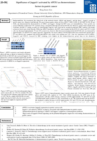
Figure 1. APEX1 expression was found in all gastric
cancer cell lines, as shown by western blot. However,
KATO-III and NCI-N87 also expressed Jagged-1. We Figure 3. Western blot of APEX1 and Jagged-1
selected two cell lines for further experiments: KATO- expression in gastric cancer cell lines (KATO-III and
III (strong expression of both proteins) and AGS (strong AGS) after APEX1 knockdown. Clear decreases in
expression of APEX1 but no Jagged-1 expression). expression were found in KATO III cells.
Figure5. Immunohistochemistry of gastric
cancer patients. Correlation between
apurinic/apyrimidinic endodeoxyribonuclease1
(APEX1), Jagged1 and CD133 expression in
gastric cancer patients. APEX1, Jagged1 and
CD133 proteins in gastric cancer patients are
shown by immunohistochemistry with anti-
APEX1, anti-Jagged1, and anti-CD133
antibodies. Brown staining indicates positive
APEX1, Jagged1, and CD133 staining (P < 0.01,
Pearson correlation test. Scale bars, 200 µm).
Figure 2. MTT assay of the chemotherapeutic drugs in
gastric cancer cells. (A: 5-FU, B: cisplatin). The cells Figure 4. MTT assay of chemotherapeutic drugs in gastric cancer cells. Cells were plated in 96-well
were plated in 96-well plates and treated with cisplatin plates and treated with cisplatin or 5-FU. After APEX1 knockdown, the MTT assay in the
or 5-FU. The IC 50 values of KATO-III cells were chemosensitive cell lines (AGS) showed a decrease in IC50 (approximately 7% and 15%) for each
higher than those of AGS cells; the values were1.5– chemotherapeutic agent (cisplatin and 5FU, respectively) (A, B). The chemoresistant cell line (KATO-
fold and 3-fold higher for cisplatin and 5-FU, III) displayed a marked decrease in IC50 (approximately 21% and 67%) for each chemotherapeutic
respectively). a g en t ( c i s p l a t i n an d 5 - FU , r es p e c t i v el y ) (C , D) .
Conclusion 1. APEX1 is activates Jagged1 expression in gastric cancer cells.
2. Jagged-1 activation by APEX1 is directly associated with chemoresistance in gastric cancer.
3. Immunohistochemical staining of APEX1 and Jagged-1 might be a chemoresponse predictor.
4. High levels of Jagged-1/Notch signaling can be potential therapeutic targets for overcoming chemoresistance in
gastric cancer.
References
1. Van Cutsem E, Haller D, Ohtsu A. The role of chemotherapy in the current treatment of gastric cancer. Gastric Cancer 2002; 5 Suppl 1:
17-22.
2. Wöhrer SS, Raderer M, Hejna M. Palliative chemotherapy for advanced gastric cancer. Ann Oncol2004; 15: 1585-1595.
3. Casaretto L, Sousa PL, Mari JJ. Chemotherapy versus support cancer treatment in advanced gastric cancer: a meta-analysis. Braz J Med
Biol Res 2006; 39: 431-440.
4. Wagner AD, Grothe W, Haerting J, Kleber G, Grothey A, Fleig WE. Chemotherapy in advanced gastric cancer: a systematic review and
meta-analysis based on aggregate data. J Clin Oncol. 2006; 24: 2903-2909.
5. Shi WJ, Gao JB. Molecular mechanisms of chemoresistance in gastric cancer. World J Gastrointest Oncol. 2016; 15; 8: 673-81.