
Page 77 - D. Cancer biology
P. 77
Phosphofructokinase 1 Platelet Isoform Promotes VEGF Expression through HIF-1α-
dependent and -independent mechanisms
Je Sun Lim and Jong-Ho Lee
Department of Health Sciences, The Graduate School of Dong-A University, Busan, 49315, Republic of Korea
BACKGROUND AIM
A. VEGF expression is regulated by transcription VEGF is overexpressed for tumor VEGF plays an important role in the angiogenesis that contributes to tumorigenesis.
vascularization, subsequent factors, such as hypoxia inducible factor-1 (HIF-1) and β-catenin
(1,2). However, it remains unclear whether the metabolic enzyme plays a role in the regulation
B. AKT activation plays a role in HIF-1α expression by increasing its translation (3). of VEGF expression. In this study, we investigated a molecular basis underlying a
C. AKT directly phosphorylates β-catenin at Ser552 (S552), which promotes nuclear glycolytic enzyme-mediated VEGF expression induced by EGFR activation in the EGFR-
translocation and transactivation of β-catenin (4).
D. In the glycolytic pathway, phosphofructokinase 1 (PFK1) catalyzes the conversion of fructose overexpressed GBM cells. Specifically, we uncovered the potential role of PFKP on the
6-phosphate and ATP to fructose-1,6-bisphosphate and ADP, which is one of the key regulatory VEGF expression.
and rate-limiting steps of glycolysis (5).
E. PFK1 exists in multiple tetrameric isozymic forms consisting of three types of subunits: METHODS
muscle (PFKM), liver (PFKL), and platelet (PFKP), and the composition of the PFK1 tetramer
varies depending on the tissue and cell type (5, 6). Cell Culture and Transfection
GBM cells including U251, EGFR-overexpressing U87 (U87/EGFR), and EGFRvIII-overexpressing U87 (U87/EGFRvIII) were were maintained in Dulbecco’s modified
Eagle’s medium (DMEM) supplemented with 10% fetal bovine serum (Capricon, USA). Cells were plated at a density of 4 × 10⁵ per 60-mm dish or 1 × 10⁵ per well
F. PFKP is the prominent PFK1 isoform in glioblastoma (GBM) cells and is overexpressed in of a 6-well plate 18 h before transfection. Transfection was performed using Polyjet transfection reagent (SignaGen) according to the manufacturer’s instructions.
DNA Constructs and Mutagenesis
human GBM specimens (7). Polymerase chain reaction (PCR)-amplified human PFKP was cloned into pcDNA3.1/hygro(+)-Flag vector. pLV/β-catenin deltaN90 (CA β-catenin) was purchased
from Addgene (Cambridge, MA). pcDNA3.1/hygro(+)-Flag PFKP Y64F was created using the QuikChange site-directed mutagenesis kit (Stratagene, La Jolla, CA).
G. Upon EGFR activation, K395-acetylated PFKP binds to EGFR, leading to EGFR-mediated pECE-Myr-HA-AKT1(delta4-129) was purchased from Addgene (Cambridge, MA). shRNA-resistant (r) PFKP contained a448c, g450c, c453t, and c456g mutations.
The following pGIPZ shRNAs were used: control shRNA oligonucleotide, GCTTCTAACACCGGAGGTCTT; PFKP
phosphorylation of PFKP Y64, which in turn binds to an SH2 domain of p85 subunit of Quantitative Real-Time PCR analysis
Total RNA isolation, reverse transcription (RT), and real-time PCR were conducted as described previously. The following primer pairs were used for quantitative
phosphoinositide 3-kinases (PI3K) and recruits PI3K to the plasma membrane. The activated real-time PCR: HIF-1α, 5′-CATAAAGTCTGCAACATGGAAGGT-3′ (forward) and 5′-ATTTAGTGGGTGAGGAATGGGTT-3′ (reverse); VEGF, 5′-
TGCAGATTATGCGGATCAAACC-3′ (forward) and 5′-TGCATTCACATTTGTTGTGCTGTAG-3′ (reverse); HPRT1, 5′-CATTATGCTGAGGATTTGGAAAGG-3′
PI3K and AKT enhances PFK1 activation and GLUT1 expression, thereby promoting aerobic (forward) and 5′-CTTGAGCACACAGAGGGCTACA-3′ (reverse).
Immunoblot Analysis
glycolysis in cancer cells and brain tumorigenesis (8). Extraction of proteins from cultured cells was performed using a lysis buffer (50 mM Tris-HCl, [pH 7.5], 0.1% SDS, 1% Triton X-100, 150 mM NaCl, 1 mM DTT, 0.5
mM EDTA, 100µM sodium orthovanadate, 100µM sodium pyrophosphate, 1 mM sodium fluoride, and proteinase inhibitor cocktail). Cell extracts were clarified via
H. PFKP Y64 phosphorylation plays an instrumental role in EGFR activation-induced β-catenin centrifugation at 13,400 g, and the supernatants (1.5 mg protein/ml) were subjected to immunoblot analysis with corresponding antibodies. Each experiment was
repeated at least three times.
transactivation through AKT activation-dependent β-catenin Ser552 phosphorylation, thereby IHC analysis and scoring
The human GBM samples and clinical information were from the Chinese Glioma Genome Atlas (CGGA, http://www.cgga.org.cn). This study was approved by the
regulating migration, invasion, and proliferation of GBM cells and brain tumor growth (9). Ethics Committee of Capital Medical University (China), and written informed consents were obtained from all patients. The tissue sections from 65 paraffin-
embedded human GBM specimens were stained with antibodies against phosphor-PFKP Y64, HIF1α, non-specific immunoglobulin as a negative control. We
I. However, the role of PFKP in the EGFR activation-induced VEGF expression of GBM cells quantitatively scored the tissue sections according to the percentage of positive cells and staining intensity, as previously defined (Ji et al., 2009). We assigned the
following proportion scores: 0 if 0% of the tumor cells showed positive staining, 0.1–1.0 if 0.1% to 1% of cells were stained, 1.1–2.0 if 1.1% to 10% stained, 2.1–3.0 if
remains unknown. 11% to 30% stained, 3.1–4.0 if 31% to 70% stained, and 4.1–5.0 if 71% to 100% stained. We rated the intensity of staining on a scale of 0 to 3: 0, negative; 1, weak;
2, moderate; and 3, strong. We then combined the proportion and intensity scores to obtain a total score (range, 0–4), as described previously (Ji et al., 2009).
Scores were compared with overall survival duration, defined as the time from the date of diagnosis to death or last known date of follow-up. The use of human
glioblastoma samples and the clinical parameters was approved by the Institutional Review Board at Capital Medical University in Beijing, Chin
RESULTS
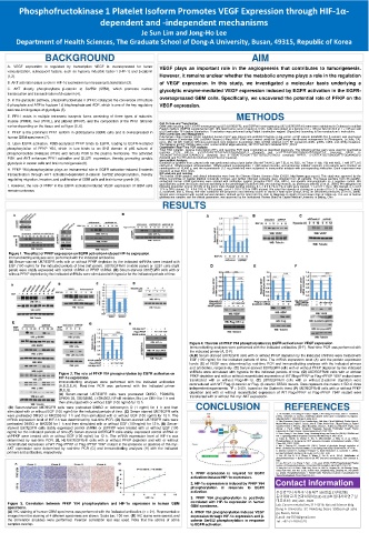
Figure 1. The effect of PFKP expression on EGFR activation-induced HIF-1α expression.
Immunoblotting analyses were performed with the indicated antibodies.
(A) Serum-starved U87/EGFR cells with or without PFKP depletion by the indicated shRNAs were treated with
EGF (100 ng/ml) for the indicated periods of time (left panel). U87/EGFRvIII (middle panel) or U251 cells (right
panel) were stably expressed with control shRNA or PFKP shRNA. (B) Serum-starved U87/EGFR cells with or
without PFKP depletion by the indicated shRNAs were stimulated with hypoxia for the indicated periods of time.
Figure 4. The role of PFKP Y64 phosphorylation by EGFR activation on VEGF expression.
Immunoblotting analyses were performed with the indicated antibodies (B-F). Real-time PCR was performed with
the indicated primer (A, D-F).
(A,B) Serum-starved U87/EGFR cells with or without PFKP depletion by the indicated shRNAs were treated with
EGF (100 ng/ml) for the indicated periods of time. The mRNA expression level (A) and the protein expression
levels (B) of VEGF were determined by real-time PCR and immunoblotting analyses with the indicated primers
and antibodies, respectively. (C) Serum-starved U87/EGFR cells with or without PFKP depletion by the indicated
Figure 2. The role of PFKP Y64 phosphorylation by EGFR activation on shRNAs were stimulated with hypoxia for the indicated periods of time. (D) U87/EGFRvIII cells with or without
PFKP depletion and with or without reconstituted expression of WT Flag-rPFKP or Flag-rPFKP Y64F mutant were
HIF-1α expression. transfected with or without Flag-HIF-1α. (E) U87/EGFRvIII cells with or without β-catenin depletion were
Immunoblotting analyses were performed with the indicated antibodies reconstituted with WT Flag-rβ-catenin or Flag- rβ-catenin S552A mutant. Data represent the means ± SD of three
(A,B,D,E,H). Real-time PCR was performed with the indicated primer independent experiments. *P < 0.001, based on the Student’s t-test. (F) U87/EGFRvIII cells with or without PFKP
(D,E,G).
(A) Serum-starved U87/EGFR cells were pretreated DMSO, PD98059, depletion and with or without reconstituted expression of WT Flag-rPFKP or Flag-rPFKP Y64F mutant were
SP600125, SB203580, LY294002, NF-κB inhibitor, Bis-I, or CKII-I for 1 h and transfected with or without HA-myr-AKT expression.
then stimulated with or without EGF (100 ng/ml) for 12 h.
(B) Serum-starved U87/EGFR cells were pretreated DMSO or Actinomycin D (1 ng/ml) for 1 h and then CONCLUSION REFERENCES
stimulated with or without EGF (100 ng/ml) for the indicated periods of time. (C) Serum-starved U87/EGFR cells
were pretreated DMSO or MK2206 for 1 h and then stimulated with or without EGF (100 ng/ml) for 12 h. The 1. J A Forsythe, B H Jiang, N V Iyer, F Agani, S W Leung, R D Koos, and G L Semenza.
(1996). Activation of vascular endothelial growth factor gene transcription by hypoxia-
mRNA expression level of HIF-1α was determined by real-time PCR. (D) Serum-starved U87/EGFR cells were inducible factor 1. Mol Cell Biol 9, 4604-4613.
2. Vijay Easwaran, Sang H. Lee, Landon Inge, Lida Guo, Cheryl Goldbeck, Evelyn Garrett,
pretreated DMSO or MK2206 for 1 h and then stimulated with or without EGF (100 ng/ml) for 12 h. (E) Serum- Marion Wiesmann, Pablo D. Garcia, John H. Fuller, Vivien Chan, Filippo Randazzo, Robert
starved U87/EGFR cells stably expressed control shRNA or shPFKP were treated with or without EGF (100 Gundel, Robert S. Warren, Jaime Escobedo, Sharon L. Aukerman, Robert N. Taylor, and
Wendy J. Fantl. (2003). β-Catenin Regulates Vascular Endothelial Growth Factor
ng/ml) for the indicated periods of time.(F) Serum-starved U87/EGFR cells stably expressed control shRNA or Expression in Colon Cancer. Cancer Research 63, 3145-3153.
3. Zhen Zhang, Li Yao, Jinhua Yang, Zhenkang Wang, and Gang Du. (2018). PI3K/Akt and
shPFKP were treated with or without EGF (100 ng/ml) for 12 h. The mRNA expression level of HIF-1α was HIF-1 signaling pathway in hypoxia-ischemia. Mol Med Rep 4. 3457-3554.
determined by real-time PCR. (G, H) U87/EGFRvIII cells with or without PFKP depletion and with or without 4. Fang D, Hawke D, Zheng Y, Xia Y, Meisenhelder J, Nika H, et al. (2007).
Phosphorylation of β-catenin by AKT promotes β-catenin transcriptional activity. J Biol
reconstituted expression of WT Flag-rPFKP or Flag-rPFKP Y64F mutant in the presence or absence of HA-myr- Chem 282, 11211-11219.
5. Mor I, Cheung EC, Vousden KH. (2011). Control of glycolysis through regulation of
AKT expression were determined by real-time PCR (G) and immunoblotting analyses (H) with the indicated PFK1: old friends and recent additions. Cold Spring Harb Symp Quant Biol 76. 211–216.
primers and antibodies, respectively. 6. Moreno-Sanchez R, Rodriguez-Enriquez S, Marin-Hernandez A, Saavedra E. (2007).
Energy metabolism in tumor cells. FEBS J 274. 1393–1418.
7. Lee JH, Liu R, Li J, Zhang C, Wang Y, Cai Q, et al. (2017). Stabilization of
phosphofructokinase 1 platelet isoform by AKT promotes tumorigenesis. Nat Commun 8.
949.
8. Lee JH, Liu R, Li J, Wang Y, Tan L, Li XJ, et al. (2018). EGFR-Phosphorylated Platelet
Isoform of Phosphofructokinase 1 Promotes PI3K Activation. Mol Cell 70. 197–210.
9. Jong-Ho Lee, Fei Shao, Jinjie Ling, Sean Lu, Rui Liu, Linyong Du, Jin Woong Chung,
Sang Seok Koh, Sun-Hee Leem, Jichun Shao, Dongming Xing, Zhiqiang An, and Zhimin
1. PFKP expression is required for EGFR Lu. (2020). Phosphofructokinase 1 Platelet Isofor Promotes β-Catenin Transactivation for
activation-induced HIF-1α expression. Tumor Development. Front. Oncol 10. 211.
2. HIF-1α expression is induced by PFKP Y64 Contact information
phosphorylation in response to EGFR
activation. 부산광역시 사하구 낙동대로 550번길 37(하단동)
3. PFKP Y64 phosphorylation is positively 동아대학교 자연과학대학(S11)-0319호암대사학연구실
대표전화 : 051) 200 - 6520
Figure 3. Correlation between PFKP Y64 phosphorylation and HIF-1α expression in human GBM correlated with HIF-1α expression in human Lab. Cancer metabolism, S11-0319, Natural Science Bldg.
GBM specimens.
specimens. Dong-A University, 37, Nakdong-Daero 550beon-gil saha-
(A) IHC staining of human GBM specimens was performed with the indicated antibodies (n = 21). Representative 4. PFKP Y64 phosphorylation induces VEGF gu, Busan, Korea
images from the staining of 4 different specimens are shown. Scale bar, 100 mm. (B) IHC stains were scored, and expression through HIF-1α expression and β- E.mail : tar7874@gmail.com
the correlation analyses were performed. Pearson correlation test was used. Note that the scores of some catenin Ser552 phosphorylation in response Tel : +82-51-200-6520
samples overlap. to EGFR activation.