
Page 73 - D. Cancer biology
P. 73
Targeting Mitochondrial Fission Factor Triggers Immunogenic Cell Death
Yu Geon Lee, Nuri Lim, Kyeong Jin Shin and Young Chan Chae
School of Life Sciences, Ulsan National Institute of Science and Technology (UNIST), Ulsan, Republic of Korea
ABSTRACT INTRODUCTION
Mitochondrial fission factor (MFF) is a molecular that control mitochondrial size and shape as Mitochondrial functions are exploited in cancer. However, how mitochondria are regulated has
well as mitochondrial dynamics that is upregulated in most cancer cells. We developed MFF- remained unclear and identification of new pathways that control mitochondrial integrity in
based cell permeable peptide (MFF peptide), which could rapidly rupture mitochondrial integrity cancer is an urgent priority. We have reported that Mitochondrial Fission Factor (MFF) as a
and induce cell death against various tumor type. In this study, we have investigated further therapeutic target in primary and metastatic cancer(1,2). Biochemically, MFF interacts with
antitumor activity of MFF peptide. Our results have revealed that treatment of MFF peptide Voltage-dependent Anion Channel (VDAC) at the mitochondrial outer membrane. Disruption of
acutely induces mitochondrial depolarization and cell death in murine tumor cells including this complex by MFF silencing or MFF peptidomimetic leads to rapid collapse of mitochondrial
B16F10 and CT26. In addition, cell death by MFF peptide exhibits the hallmarks of functions with increased mitochondrial outer membrane permeability, release of apoptogenic
immunogenic cell death including secretion of high mobility group box1 (HMGB1) and
adenosine triphosphate (ATP), and translocation of calreticulin to the cells surface. Moreover, factors, and activation of cell death pathway. In addition, treatment of MFF peptide with cancer
cells induced acutely focal necrosis that was accompanied by extracellular release of HMGB1.
mice treated with MFF peptide results in increased immune response in blood serum and tumor Therefore, we have further questioned the precise role and mechanism of cell death by MFF
tissue with tumor size reduction. These finding suggests that therapeutic targeting MFF may targeting.
provide actionable strategy to impair cancer development and progression.
RESULTS
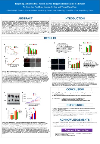
Figure 1. MFF peptidomimetic induces mitochondrial cell death in murine cancer cells. Figure 2. MFF peptidomimetic elicits hallmarks of immunogenic cell death.
(A and B) Murine cancer cells (Ct26 and B16F10) were treated with increasing concentration (0-50 μM) of cell- (A-E) Murine cancer cells (Ct26 and B16F10) were treated for 2 hours with indicated concentration (0-20 μM) of
permeable MFF peptide and analyzed for 1 hr. (A) Cell growth was analyzed by direct cell counting. (B) The cell cell-permeable MFF peptide. (A) Release of high mobility group box 1 (HMGB1) from cells exposed to MFF
number was calculated by the trypan blue exclusion using automated cell counter (Countess II FL, Invitrogen). peptide. Culture supernatant or pellet were collected, and proteins were analyzed by Western blotting. (B)
Percentage of dead cells was calculated using the following formula: [100 – (number of live cells/number of total Release of ATP from cells exposed to MFF peptide. Culture supernatant were collected, and extracellular ATP
cells × 100)]. The data are expressed as the Mean±SD from two independent experiments. (C and D) Ct26 and was measured by Bioluminescence detection system (Promega, FF2000). The data are expressed as the
B16F10 cells were treated with indicated concentration (10 or 20 μM) of cell-permeable MFF peptide and analyzed Mean±SD from two independent experiments. (C-E) Translocation of calreticulin (CRT) to surface of cells
for mitochondrial membrane potential by TMRE labeling. FCCP (5 μM) was used as negative control (C) Change in exposed to MFF peptide. (C-D) Ct26 and B16F10 cells were transfected with CRT-GFP and cultured in the
TMRE labeling were quantified by fluorescence microplate reader (Ex/Em=535/595 nm). (D) TMRE fluorescence indicated conditions with cell-permeable MFF peptide. (C) Cells were fixed and counterstained with the
reactivity was imaged by fluorescence microscopy. TMRE, tetramethylrhodamine, ethyl ester; DIC, differential chromatin dye Hoechst 33342 and subjected to fluorescence microscopy and subjected to fluorescence
interference contrast. Scr, cell-permeable scrambled peptide; MFF, cell-permeable MFF peptide. microscopy (representative microphotographs in C and statistical analyses in D). The data are expressed as the
Mean±SD from two independent experiments. (E) Culture cells were harvested, and proteins were analyzed by
Western blotting. Scr, cell-permeable scrambled peptide; MFF, cell-permeable MFF peptide.
CONCLUSION
▶ Targeting MFF using peptidomimetic agent disrupts mitochondrial integrity and exerts anticancer
activity in cancer cells.
▶ Mitochondrial cell death induced by MFF targeting can induce immunogenic cell death.
▶ Ongoing study: 1. T-cell activation or infiltration by MFF peptidomimetic
2. Activation of DC function by MFF peptidomimetic
3. Alteration of immune checkpoint activity by MFF peptidomimetic
REFERENCES
1. Seo et al. Mitochondrial fission factor is a novel Myc-dependent regulator of mitochondrial permeability
in cancer. EBioMedicine (2019)
2. Seo et al. MFF Regulation of Mitochondrial Cell Death Is a Therapeutic Target in Cancer.
Cancer Research (2019)
3. Zhou et. al. The oncolytic peptide LTX-315 triggers immunogenic cell death. Cell Death and Disease (2016)
4. Hossain et al. Dinaciclib induces immunogenic cell death and enhances anti-PD1–mediated tumor
suppression. The Journal of Clinical Investigation (2018)
ACKNOWLEDGEMENTS
Figure 3. MFF peptidomimetic inhibits in vivo tumor proliferation and stimulates immune response. We would like to thank members of the Chae laboratory for their support, collegiality, and critical review of the
(A and B) Inhibition of tumor growth by MFF peptide in vivo. B16F10 cells (1x10 6 cells in HBSS) were engrafted manuscript.
onto the flanks of immunocompetent mice, and animals randomized in two groups were treated with of cell- This work was supported by the National Research Foundation of Korea grant funded by the
permeable scrambled peptide or MFF (50 mg/kg, daily i.p.) with quantification of tumor growth. The data are Ministry of Education (2019R1I1A1A01056609)
expressed as the Mean±SD (n=4). **p=0.0115. Differences between the control versus MFF peptide treatment
groups are shown as tumor images (A) and tumor sizes (B). (C and D) Induction of immune response by MFF
peptide. (C) Total RNA of tumor tissue was isolated and indicated mRNA levels were measured by qRT-PCR. The
data are expressed as the Mean±SD (n=3) ***p<0.0001. (D) Blood were collected, and serum was isolated. Levels Contact information
of serum cytokines (IL-1β and IL-6) were quantified by ELISA kit analysis (R&D System). The data are expressed as
the Mean±SD (n=3). **p=0.0057; ***p=0.0006. Scr, cell-permeable scrambled peptide; MFF, cell-permeable MFF School of Life Sciences, Ulsan National Institute of Science and Technology, UNIST-gil 50
peptide.
Ulsan 44919, Republic of Korea
Tel. +82-52-217-2629; Email: ugun2@unist.ac.kr