
Page 71 - D. Cancer biology
P. 71
Targeting MFF Induces the Mitochondrial Cell Death via Dissociation of the Anti-apoptotic Regulator
BCL-XL-VDAC Complex in Cancer
Yu Geon Lee, Nuri Lim and Young Chan Chae
School of Life Sciences, Ulsan National Institute of Science and Technology (UNIST), Ulsan, Republic of Korea
ABSTRACT
Mitochondria are hubs of multiple cell death pathway including apoptosis and necrosis. These processes mostly involve a sudden increase in the mitochondrial outer membrane permeability
(MOMP). However, how modulators of mitochondrial cell death are programmed in cancer to promote their survival has not been fully understood. In our previous study, we identified that
mitochondrial fission factor (MFF) formed complex with the voltage-dependent anion channel (VDAC), a key modulator of MOMP. Here, we have shown that MFF forms heterotrimeric complex with
VDAC and anti-apoptotic BCL-XL protein on mitochondrial outer membrane (MOM). This complex is required for cell death mechanism including release of apoptogenic cytochrome c from
mitochondria into cytosol. Accordingly, MFF regulation to modulate interaction of VDAC with BCL-XL proteins is sufficient to induce mitochondrial cell death with loss of mitochondrial membrane
potential. This finding indicates that mitochondrial fission can be exploited to couple to the machinery of modulator of mitochondrial cell death including antiapoptotic VDAC-BCL-XL complex to
improve tumor survival.
INTRODUCTION
Evading cell death signaling is a hallmark of most cancer and contributes to chemo-resistance. Apoptotic cell death is regulated by the BCL-2 family which includes pro- and anti-apoptotic
subgroups(1). Therefore, cancer cells have straightforward strategy for survival to evade programmed cell death by upregulating anti-apoptotic BCL-2 members including BCL-2, BCL-XL, and MCL-
1 (Picture A)(1). In addition, BCL-2 family proteins regulates MOMP by forming BAX/BAK oligomeric pore, thereby inducing cell death signaling. In this context, VDAC channels have been reported
to influence cell death by interacting with BCL-2 family proteins (BCL-XL, BAX, and BAK) on MOM (Picture B)(2). Although the precise determinants of their recruitment to the MOM to mediate cell
death are unclear, many chemotherapeutic agent, called BH3 mimetics, have attempted to target BCL-2 proteins indirectly or directly to drive cell death. Unfortunately, there have been no trials for
drugs targeting BCL-2 family that effectively act on solid tumor, which is thought to be because we have not fully understood their exact interaction mechanism on MOM(3). Recently, we identified
MFF and VDAC make homo- and heterodimeric complexes, which is play a crucial role in regulating MOMP. Furthermore, disruption of MFF-VDAC complex triggered cell death with depolarized
mitochondria(4,5). However, mechanistic link between mitochondrial fission factors and death mediators in mitochondria in cancer has not been fully elucidated. In this study, we have uncovered an
interplay of MFF with VDAC-BCL-XL complex that can be a therapeutic actionable target by modulating mitochondrial cell death in cancer.
RESULTS
Figure 3. Pro-survival role of MFF-VDAC-BCL-XL complex in cancer. (A-C) PC3 cells were
transfected with Flag-BCL-XL with or without GFP-MFF for 48 hr. (A) Cells were treated with
H 2 O 2 (100 μM) for 2 hr and fractionated into cytosolic (Cyto) and mitochondrial (Mito) extracts and
analyzed by western blotting. Cyt, cytochrome. (B) Cells were treated with increasing
concentration (0-10 μM) of CaCl 2 for 1 hr and analyzed for mitochondrial membrane potential by
TMRE labeling. Change in TMRE labeling were quantified by fluorescence microplate reader
(Ex/Em=535/595 nm). Mean±SD (n=3). ***p=0.0002. (C) Cells were treated with staurosporine
(STS, 0.1 μM), doxorubicin (DOX, 1 μM), or cisplatin (CIS, 20 μM) for 24 hr. Cell growth was
analyzed by direct cell counting. Mean±SD (n=3). *p=0.0312; **p=0.0063; ***p=0.0049. (D and E)
PC3 cells were seeded in plates and treated with MFF peptide (5 μM) with or without inhibitor for
BCL-XL (5 μM). Colony formation was assessed by crystal violet staining after 14 days and was
quantified (E). Mean±SD (n=3). ***p=0.0001. (F and G) DU145 cells were allowed to form
Figure 1. Mitochondrial MFF-VDAC-BCL-XL complex in cancer. (A) Heatmap of proteomics identification of spheroids over 4 days and treated with MFF peptide (5 μM) with or without inhibitor for BCL-XL (5
mitochondrial proteins co-precipitated with a Flag-MFF or control vector from PC3 cell lysates. A fold enrichment in μM). (F) Representative microscopy images of spheroid formation after treatment of MFF peptide
spectral count (left) and relative spectral count (right) are shown. (B) Identified proteins are classified by Gene with or without inhibitor of BCL-XL (scale bar: 400 μm). (G) Size of tumor spheroid in each group
Ontology enrichment analysis (DAVID, NIH). (C) Co-expression analysis of identified proteins with MFF in human was analyzed using image J software. Mean±SD (n=50). ***p<0.0001. XL inhibitor, inhibitor of
prostate cancer dataset (TCGA). (D) Co-expression analysis for MFF with death mediator of mitochondrial (VDACs BCL-XL (A1331852).
and BCL-2 family) in various cancer database (TCGA). (E) The protein levels of MFF and anti-apoptotic BCL-2 family
in the indicated cell lines were analyzed by western blotting. (F) PC3 cells were transfected with control vector, Flag-
MFF and were immunoprecipitated with anti-Flag antibody and analyzed by western blotting. (G) PC3 cells were CONCLUSION
transfected with control vector, Flag-BCL-XL and were immunoprecipitated with anti-Flag antibody and analyzed by
western blotting. (H) PC3 cells were transfected with non-targeting siRNA (siCtrl) or MFF-directed siRNA (siMFF) and
transfected with Flag-VDAC were immunoprecipitated with anti-Flag antibody and analyzed by western blotting. (I) - We have shown that MFF-VDAC-BCL-XL complex as promising regulator of
PC3 cells were transfected with siCtrl or siMFF and transfected with Flag-BCL-XL were immunoprecipitated with anti- mitochondrial cell death in cancer.
Flag antibody and analyzed by western blotting. TCE, total cell extract. - Targeting MFF can be therapeutic strategy to overcome resistance to anti-
apoptotic BCL-2 proteins including BCL-XL.
- Destruction of the MFF-VDAC-BCL-XL complex may provide therapeutic window
in malignant tumors and open the strategy to bypass the drug resistance
mechanisms maintained by the anti-apoptotic Bcl2 proteins.
REFERENCES
1. Czabotar et al. Control of apoptosis by the BCL-2 protein family: implications for physiology and therapy.
Nature Reviews Molecular Cell Biology (2013).
2. Tsujimoto et al. VDAC regulation by the BCL-2 family of proteins. Cell Death and Differentiation (2000).
3. Warren et al. BCL-2 family isoforms in apoptosis and cancer. Cell Death and Disease (2019).
4. Seo et al. Mitochondrial fission factor is a novel Myc-dependent regulator of mitochondrial permeability in
cancer. EBioMedicine (2019).
5. Seo et al. MFF Regulation of Mitochondrial Cell Death Is a Therapeutic Target in Cancer.
Cancer Research (2019).
ACKNOWLEDGEMENTS
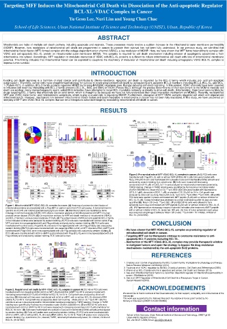
Figure 2. Regulation of cell death by MFF-VDAC-BCL-XL complex in cancer (A) DU145 or PC3 cells were
transfected with non-targeting siRNA (siCtrl) or MFF-directed siRNA (siMFF) for 48 hr and were treated with We would like to thank members of the Chae laboratory for their support, collegiality, and critical review of the
increasing concentration (0-10 μM) of inhibitor for BCL-XL for 24 hr. Cell growth was analyzed by direct cell manuscript.
counting. (B) Indicated cell lines were transfected with siCtrl or siMFF, with or without BCL-XL-directed siRNA This work was supported by the National Research Foundation of Korea grant funded by the
(siBCL-XL) for 48 hr. Cell growth was analyzed by direct cell counting. . Mean±SD (n=3). ***p<0.001. (C) The Ministry of Education (2019R1I1A1A01056609)
conditions are as in A and analyzed for mitochondrial membrane potential by TMRE labeling. Change in TMRE
labeling were quantified by fluorescence microplate reader (Ex/Em=535/595 nm). (D and E) DU145 cells were
transfected with siCtrl or siMFF for 48 hr and were treated with increasing concentration (0-10 μM) of inhibitor for Contact information
BCL-XL for 24 hr. (D) Cells were fractionated into cytosolic (Cyto) and mitochondrial (Mito) extracts and analyzed
by western blotting. (E) Total cell lysates were analyzed by western blotting. (F) PC3 cells were transfected with
siCtrl or siMFF, with or without BCL-XL-directed siRNA (siBCL-XL) for 48 hr. Total cell lysates were analyzed by School of Life Sciences, Ulsan National Institute of Science and Technology, UNIST-gil 50
western blotting. Cyt, cytochrome; Clv, cleaved; TMRE, tetramethylrhodamine ethyl ester; XL inhibitor, inhibitor of Ulsan 44919, Republic of Korea
BCL-XL (A1331852). Tel. +82-52-217-2629; Email: ugun2@unist.ac.kr