


Page 211 - ebook
P. 211
F-actin-mediated mechanosensation controls
STAT6-dependent M2 macrophage activation
Jeong-Ki Kim and Dong-Hwee Kim 1, 2
1
Applied Mechanobiology Group
1 KU-KIST Graduate School of Converging Science and Technology, Korea University, Seoul, South Korea
2 Department of Integrative Energy Engineering, College of Engineering, Korea University, Seoul, South Korea
Abstract
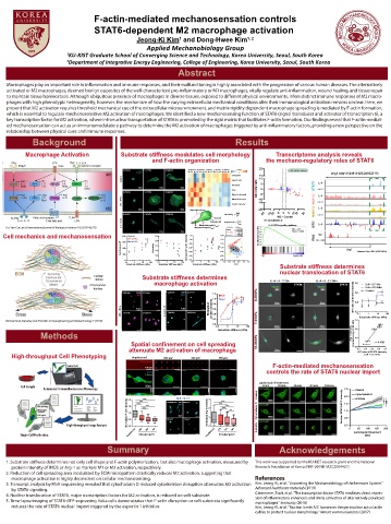
Macrophages play an important role in in ammation and immune responses, and their malfunctioning is highly associated with the progression of various human diseases. The alternatively
activated or M2 macrophages, deemed benign opposites of the well-characterized pro-in ammatory or M1 macrophages, vitally regulate anti-in ammation, wound healing, and tissue repair
to maintain tissue homeostasis. Although ubiquitous presence of macrophages in diverse tissues, exposed to di erent physical environments, infers distinct immune responses of M2 macro-
phages with high phenotypic heterogeneity, however, the mechanism of how the varying extracellular mechanical conditions alter their immunological activation remains unclear. Here, we
present that M2 activation requires threshold mechanical cue of the extracellular microenvironment, and matrix rigidity dependent macrophage spreading is mediated by F-actin formation,
which is essential to regulate mechanosensitive M2 activation of macrophages. We identi ed a new mechanosensing function of STAT6 (signal transducer and activator of transcription 6), a
key transcription factor for M2 activation, where intranuclear transportation of STAT6 is promoted by the rigid matrix that facilitates F-actin formation. Our ndings reveal that F-actin-mediat-
ed mechanosensation can act as an immunomodulatory pathway to determine the M2 activation of macrophages triggered by anti-in ammatory factors, providing a new perspective on the
relationship between physical cues and immune responses.
Background Results
Macrophage Activation Substrate stiffness modulates cell morphology Transcriptome analysis reveals
and F-actin organization the mechano-regulatory roles of STAT6
20
0.20 kPa DAPI 7.44 kPa 34.88 kPa 34.88 kPa 23.43 kPa 7.44 kPa 1.00 kPa 0.20 kPa z-score M2 signature genes
F-Actin M0 M1 M2 M0 M1 M2 M0 M1 M2 M0 M1 M2 M0 M1 M2 -2 0 Arg1 (chr10:24914125-24932715)
Circularity 10
10
Aspect ratio
Perimeter 5 IL-4
20 m 20 m 20 m STAT6
Area CD + M2 / M2 0 STAT6 cont
0.20 kPa 7.44 kPa 34.88 kPa
MajorAxisLength -5 Arg1 IL-4
MinorAxisLength -10 RNAPII-S2
-10 cont
F-actin intensity
20 m 20 m 20 m -20 IL-4
Equivalent ellipse
-20 -10 -10 -5 0 5 10 10 20 RNAPII-S5
0.20 kPa 7.44 kPa 34.88 kPa Circularity cont
Area (A) M2 / Contol
L CD: Cytochalasin D
Perimeter (P) Aspect ratio L minor
MinorAxisLength (L minor ) major IL-4-STAT6 signaling pathway GRO IL-4
Liu, Yan-Cun, et al. International journal of biological sciences 10.5 (2014): 520 MajorAxisLength (L major ) Activation Sti ness 0.7
Control 34.88 kPa 0.6 NES = 2.67
20 m 20 m 20 m LPS + IFN-γ 23.43 kPa 0.5 Nom. P < 0.001
7.44 kPa
Cell mechanics and mechanosensation 1200 Control 0.9 1.2 0.6 IL-4 + IL-13 1.00 kPa Enrichment score (ES) 0.4 RNA CD + M2
M2
0.20 kPa
0.3
LPS + IFN-γ
0.4
1.0
0.2
0.8
IL-4 + IL-13
900
Cell Area (μm 2 ) 600 Circularity 0.7 Normalized F-actin intensity 0.8 PC2 -0.2 0.20 kPa 0.0 Arg1 Datasets from GEO (GSE10670)
0.1
0.2
0.0
0.6
0.6
0.4
300
-0.6
0.0
0.4
0 0.5 0.2 -0.4 M2 CytoD + M2
0.1 1 10 100 0.1 1 10 100 0.1 1 10 100 -1.0 -0.5 0.0 0.5 1.0 1.5
Substrate Stiffness (kPa) Substrate Stiffness (kPa) Substrate Stiffness (kPa) PC1 Substrate stiffness determines
nuclear translocation of STAT6
Substrate stiffness determines
IL-4 + IL-13 2hr
IL-4 + IL-13 24hr
macrophage activation Nucleus STAT6 Nucleus STAT6 0.8 IL4 + IL13 2hr
STAT6 STAT6 IL4 + IL13 24hr
DIC DIC Control
0.20 kPa DAPI 3.15 kPa 0.20 kPa DAPI 3.15 kPa 1.2 0.6
iNOS Arg-1 iNOS
1.0 Arg-1 20 m 20 m N/T ratio of STAT6 intensity 0.4
Normalized protein intensity
0.6
50 m 50 m 0.8
7.44 kPa 34.88 kPa 7.44 kPa 34.88 kPa 0.4 0.2 1 10 100
0.1
Mohammed, Danahe, et al. Frontiers in bioengineering and biotechnology 7 (2019) 0.2 1.2 Substrate stiffness (kPa)
0.0
0.1 1 10 100 20 m 20 m 0.8
Substrate stiffness (kPa)
Methods Arg-1 intensity (normalized by 34.88 kPa) 0.4
Spatial confinement on cell spreading r = 0.9725
attenuate M2 activation of macrophage 20 m 0.0 0.3 0.4 0.5 0.6 0.7
N/T ratio of STAT6 intensity
High-throughput Cell Phenotyping Unpatterned 800 m 2 300 m 2 150 m 2 (IL4 + IL13 2hr)
Fibronectin
Stained Cells F-actin-mediated mechanosensation
100 m
DAPI controls the rate of STAT6 nuclear import
F-Actin
Leptomycin B treatment
Cell Sample 20 m 0 min 30 min 60 min 120 min 180 min
Automated Immuno uorescence Microscopy STAT6-GFP 2.5 Control
10 m Cytochalasin D
2.0 *** IL-4 + IL-13 3 Control 20 m 2.0 0.20 kPa
*** Unpatterned DAPI 800 m 2 *** *
*** Arg-1 *** STAT6-GFP 1.5
**
Morphology 1.5 *** 2 *** *** Nuclear STAT6-GFP intensity
Molecular *** ** 1.0
content Normalized F-actin intensity 1.0 Normalized Arg-1 intensity Cytochalasin D
Cell cycle 50 m
1 20 m 0.5
0.5 300 m 2 150 m 2
STAT6-GFP
High-throughput Image Analysis 0.0 0 0.20 kPa 0.0 0 50 100 150 200
Unpatterned 800 300 150 Unpatterned 800 300 150 20 m Leptomycin B treatment
Single-Cell Pro le data Circular ( m ) Circular ( m ) (min)
2
2
Summary Acknowledgements
1. Substrate sti ness determines not only cell shape and F-actin polymerization, but also macrophage activation, measured by This work was supported by the KU-KIST research grant and the National
protein intensity of iNOS or Arg-1 as markers M1 or M2 activation, respectively. Research Foundation of Korea (NRF-2019R1A2C2004437)
2. Reduction of cell spreading area modulated by ECM micropattern drastically reduces M2 activation, suggesting that
macrophage activation is highly dependent on cellular mechanosensing. References
3. Transcript analysis by RNA sequencing revealed that cytochalasin D-induced cytoskeleton disruption attenuates M2 activation Kim, Jeong-Ki, et al. "Unraveling the Mechanobiology of the Immune System."
by STAT6 signaling. Advanced healthcare materials (2019)
4. Nuclear translocation of STAT6, major transcription factors for M2 activation, is reduced on soft substrate. Czimmerer, Zsolt, et al. "The transcription factor STAT6 mediates direct repres-
sion of in ammatory enhancers and limits activation of alternatively polarized
5. Time-lapse imaging of STAT6-GFP-expressing HeLa cells demonstrates that F-actin disruption or soft substrate signi cantly macrophages." Immunity (2018)
reduces the rate of STAT6 nuclear import triggered by the exportin 1 inhibitor. Kim, Jeong-Ki, et al. "Nuclear lamin A/C harnesses the perinuclear apical actin
cables to protect nuclear morphology." Nature communications (2017)

