


Page 181 - ebook
P. 181
Poster Title
Ovarian Cancer Stem cells targeted anti-CD44 conjugated, and cisplatin loaded PLGA nanoparticles on
tumor associated macrophages embedded 3D spheroid models
ㅇ ㅇ ㅇ ㅇ
Researchers’/Presenters’ Names
Institution/Organization/Company
Shrestha Samjhana , Simmyung Yook * 1
1
1 College of pharmacy, Keimyung University, Daegu, 42601, Republic of Korea
Abstract Results
Background: The presence of cancer stem cells (CSCs) and their interplay with tumor associated Characterization of Cis-loaded PLGA nanoparticles
macrophage (TAM) have made ovarian cancer (OC) drug resistant toward Cisplatin (Cis). CSC surface
0.6
marker CD44 targeted therapy using PLGA nanoparticles can be an attractive therapeutic strategy for y = 0.0006x - 0.0176 Average size of PLGA NPs 119.8 nm ± 1.84 nm
treatment of recurrent OC. In this study, since 3D culture spheroids models can mimic the OC tumor 0.5 R² = 0.997
microenvironment, we developed 3D cancer model in the presence of TAM and evaluated the effect of 0.4 Loading capacity (LC) of Cis on PLGA 5.55 % ± 0.12%
CD44 targeted and Cis-loaded PLGA nanoparticles (anti-CD44-Cis-PLGA NPs) for treatment of recurrent Abosrbance 0.3 NPs
OC. Encapsulation efficiency (EE) of Cis on 18.5% ± 0.41%
0.2 PLGA NPs
Methods: Cis was loaded in PLGA with double emulsion solvent evaporation procedure. The formulation
0.1
is characterized by average particle size, zeta potential and polydispersity (PDI), loading capacity (LC) Conjugation efficiency (CE) of anti- 61.73% ± 10.1 %
0 CD44 on PLGA NPs
and encapsulation efficiency (EE). The surface modification of Cis-loaded PLGA was done by EDC/NHS 0 200 400 600 800 1000 1200
Concentration (ug/ml)
chemistry and is characterized by pierce bichinchoninic acid (BCA) protein assay.
Fig 4: Calibration curve of Cis Table 1: Characteristics of Cis loaded PLGA nanoparticles
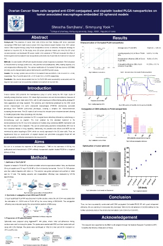
Results: The average particle size and PDI of Cis-loaded PLGA were119.8 ±1.84 nm and 0.14 ± 0.02,
respectively. The LC and EE were 5.55 ± 0.12 % and 18.5 ± 0.417% respectively.
6
Conclusion: Our results demonstrated that the Cis-PLGA NPs were successfully constructed, and this
targeted nano system can be a potential candidate for treatment of recurrent OC.
4
Introduction Loading capacity % 2
Ovarian cancer (OC) presents the heterogenous group of tumor, being the fifth major cause of
0
mortality among American women. OC shows high recurrence rate and chemoresistance because of 10% 20% 30%
the presence of cancer stem cells (CSC), with its self-renewing and differentiating ability, making OC Cisplatin concentration
more aggressive and drug resistant. The cytokines and chemokines produced by the CSC could
Fig 5: Loading capacity of Cis
convert macrophages into tumor associated macrophages (TAM-M2, alternatively activated) Fig 6: FTIR spectra of PLGA NPs, free Cis and Cis-PLGA NPs.
phenotype from TAM-M1 (anti-tumor) phenotype, creating a complex OC microenvironment Conjugation of CD44 antibody on PLGA nanoparticles
increasing the stemness. The 3D model of cell mimics this intricate tumor microenvironment allowing
0.15 80
to showing appropriate tissue physiology and structure.
0.14 y = 0.0378x + 0.064
The standard management procedure for OC is surgical tumor debulking followed by radiotherapy or 0.13 R² = 0.9998 60
chemotherapy such as cisplatin. The main problem for this standard treatment is the 0.12
chemoresistance by the OC due to the presence of multiple drug resistance (MDR) mechanism and Absorbance 0.11 Conjugation efficiency % 40
the short circulation time of cisplatin. This can be solved using polymeric nanoparticles (NP) due to 0.1
0.09
their sustained release of drug capability. Additionally, the possible side effects and MDR can be 0.08 20
minimized by active targeting to CD44, which are overly expressed in the OC stem cells. Thus, we 0.07
0
hypothesized that our construction of cisplatin loaded and anti-CD44 conjugated PLGA NP will 0.06
0 0.5 1 1.5 2 2.5 1 10 100 200
provide possibility of effective treatment for the recurrent OC. Concentration (ug/ul) Antibody(Ab) concentration (ug/ml)
Aims Fig 7: Calibration curve of BCA Fig 8: Antibody conjugation efficiency
Our aim is to evaluate the response of M2 phenotypic – TAM on the stemness of OC by the Optimization of tumor spheroid
synthesized and characterized a CD44 antibody incorporated cisplatin loaded PLGA for a targeted
delivery of drug.
Methods
1. Synthesis of Cis-PLGA NP
Cisplatin is loaded in PLGA NP by double emulsion solvent evaporation method. Here, we dissolved
30 mg of cisplatin and 70 mg of PLGA NP in 20 ml of dimethyl formamide. 15 ml of 1% PVA solution
was then added dropwise with stirrer on. The solution was probe sonicated and purified at 12000
rpm for 12 min. The loading capacity and encapsulation efficiency was measured by UV-Vis
spectroscopy.
Fig 9: Optimization of cell density for 3D-spheroid Fig 10: Spheroid diameter
Fig 1: Schematic representation of Cis loaded PLGA
2. Synthesis of conjugation of anti-CD44 and PLGA NP Conclusion
2 mg of lyophilized PLGA NP was dissolved in buffer solution, where the anti-CD44 was added after
the stimulation of -COOH end of PLGA NP by the cross linking of EDC/NHS. The conjugation
efficiency was evaluated against the concentration gradient of BSA assay. Thus, we have successfully constructed anti-CD44 conjugated Cis-loaded PLGA NP with good entrapment
efficiency. We are planning to incorporate M2 phenotypic TAM into the 3D spheroid of SKOV3 cell line for the
further cytotoxicity study of the constructed formulation (anti-CD44-Cis-PLGA NP).
Fig 2: Schematic illustration of antibody conjugation Acknowledgement
3. Preparation of 3D spheroid model
Spheroids were prepared using Aggrewell TM 400 plates, where 100ul anti-adherence rinsing
solution was used for rinsing the plates. The plates were washed with PBS and then loaded with cell This research was supported by the BK21 fourth program through the National Research Foundation (NRF)
along with 2.5% Matrigel. The plates were centrifuged at 100g for 3 min and left for incubation at funded by the Ministry of Education of Korea.
37°C and 5% CO2.
Fig 3: Schematic presentation of 3D spheroid

