


Page 179 - ebook
P. 179
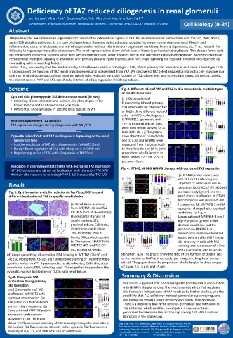
Deficiency of TAZ reduced ciliogenesis in renal glomeruli

                               1
                      1
              Jae Hee Jun , Minah Park , Gyuyeong Rha, Yejin Ahn, Jin-ui Min, Jong Hoon Park 1,*
              1 Department of Biological Science, Sookmyung Women’s University, Seoul, 04310; Republic of Korea  Cell Biology (B-24)
     Abstract
     The primary cilia are antenna-like organelles and transmit the extracellular signals to cell that maintain cellular homeostasis with the Shh, Wnt, Notch,
     and mTOR signaling pathways. In the case of ciliary defect, there are various diseases polydactyly, sensorineural deafness, renal fibrosis and
     inflammation, cystic renal disease, and retinal degeneration without cilia in sensory organs such as kidney, brain, and pancreas, etc. Thus, research for
     following to regulate primary cilia is important. The most representative ciliary defect case in kidney is polycystic kidney disease. This disease forms cysts
     full of fluid and loses renal function along with various complications, which requires continuous dialysis or kidney transplantation. Recent studies have
     revealed that the hippo signaling is associated with primary cilia and cystic diseases, and YAP, hippo signaling key regulator, involved in ciliogenesis by
     modulating actin remodeling factors.
      In this study, we investigated the mechanism by TAZ deficiency which is orthologous YAP, affects primary cilia formation in each renal tubule type. Little
     is known about the function of TAZ regulating ciliogenesis and independent from YAP. We discovered TAZ defect reduced primary cilia only in glomerular
     cells not renal collecting duct cells or proximal tubule cells. Although our study focused on TAZ, ciliogenesis, and other ciliary genes, the results suggest
     the distinct roles of YAP and TAZ, specifically in terms of ciliary regulation in kidneys tubules.
     Scheme                                              Fig. 3. Different roles of YAP and TAZ in cilia formation in multiple types
                                                         of renal tubular cells
      Cyst and Cilia phenotypes in TAZ defect mouse model (in vivo)  (a-l) Observations of
      • Screening of cyst formation and primary cilia phenotypes in Taz-  fluorescently labeled primary
       floxed:KSP-cre and Taz-floxed:HoxB7-cre mice.      cilia after silencing of either YAP
      • Differential TAZ expression in  specific renal tubules in WT
                                                          or TAZ in three different types of
                                                          cells—mIMCD, collecting duct;
      Relationship between TAZ and cilia                  SV40MES13, glomeruli; and
      TAZ expression changes during ciliogenesis with NIH/3T3   TKPTS, proximal tubule. Cells
                                                          were then serum starved for at
                                                          least 24 h. (b, f, j) The graphs
      Opposite roles of YAP and TAZ in ciliogenesis depending on the renal   show the ratio of ciliated cells
       tubular cell type                                  and (c, g, k) cilia lengths were
      • Positive regulation of TAZ with ciliogenesis in SV40MES13 cell.  measured from the basal body
      • No significant regulation of TAZ with ciliogenesis in IMCD cell.  to the ciliary tip and (d, h, l) the
      • Negative regulation of TAZ with ciliogenesis in TKPTS cell.  proportions of cilia lengths in
                                                          three ranges: <2.5 µm, 2.5 - 5
                                                          μm, and >5 μm.
      Validation of ciliary genes that change with decreased TAZ expression  Fig. 4. IFT140, NPHP6, NPHP9 changed with decreased TAZ expression
      •IFT140  decrease and abnormal localization with cilia under TAZ  K/D
      •Primary cilia recovery by reducing NPHP 6 & 9 increased by TAZ K/D           (a) IFT140 protein expression
                                                                                   with YAP or TAZ silencing and
     Result                                                                        subjected to 24 hours of serum
                                                                                   starvation. (b, C) ICC of IFT140 (red)
     Fig. 1. Cyst formation and cilia reduction in Taz-floxed:KSP-cre and          and basal body (green) and the
     different localization of TAZ in specific renal tubules                       graph shows localization of IFT140
                                                                                   in primary cilia was classified into
                                  (a) Renal tissue sections                        4 categories. (d) NPHP6 & 9 mRNA
                                  from WT, TAZ cKO and TAZ-                        expression changed with identical
                                  CD cKO) mice at 36 weeks (b)                     conditions. (e, f, g, h)
                                  Fluorescence staining of                         Accumulation of NPHP6 & 9 (red)
                                  tubule markers. LTL,                             in primary cilia (green) under
                                  proximal tubule; Calbindin,                      identical conditions and the
                                  distal convoluted tubule;                        graphs show NPHP 6 & 9
                                  THP, ascending loop of                           fluorescence intensities localized
                                  Henle; DBA, collecting duct.                     around primary cilia. (i-l) Primary
                                  (c) The ratio of 2KW/TBW in                      cilia recovery in cells with TAZ
                                  WT, TAZ cKO, and TAZ CD-                         silencing and knockdown of either
                                  cKO mice at 36 weeks.                            NPHP 6 or 9 after 24 h of serum
    (d) Graph quantifying cilia-positive DBA labeling in WT, TAZ CD-cKO and   starvation. (j, k) The graphs show the ratio of the number of ciliated cells
    TAZ cKO mouse renal tissues. (e) Fluorescence staining of TAZ with tubule-  to the number of DAPI-stained nuclei per image and lengths of primary
    specific markers in WT.  Synaptopodin, renal podocytes; Calbindin, distal   cilia. (l) The graphs show the proportions of cilia lengths in three ranges:
    convoluted tubule; DBA, collecting duct. The magnified images show the   <2.5 μm, 2.5 - 5 μm, and >5 μm.
    cytosolic/nuclear localization of TAZ in each renal tubule.
                                                          Summary & Discussion
     Fig. 2. Changes in TAZ
     localization during primary                          Our results suggested that TAZ may regulate primary cilia in cooperation
     cilia formation.                                     with NPHP in the glomerulus. The mechanism by which TAZ regulates
     (a-d) Observations of TAZ                            cilia formation independent of YAP needs to be further studied, and the
     localization in NIH3T3 cells                         possibility that TAZ deficiency-induced glomerular cysts may regulate
     upon serum starvation. (a)                           cyst formation through ciliary recovery also needs to be discussed.
     Acetylated α-tubulin-labeled                         There is a possibility that NPHP restores glomerular cyst formation in
     primary ciliary axoneme. (b)                         TAZ cKO mice, which could be investigated if experiments are
     Comparison of YAP/TAZ protein                        performed to determine the relationship among TAZ, NPHP and cyst
     expression under serum-                              formation in the glomerulus.
     starvation. (c, d) The graph
     shows the fluorescence intensity of TAZ around primary cilia  and ratio of   Jong Hoon Park, Ph.D. Professor, Department of Biological Science Director,
     the nuclear TAZ fluorescence intensity to the cytosolic TAZ fluorescence   Post Genome Research Center & Research Certer for Molecular Medicine,
                                                        Sookmyung Women's University, 100 Changpa-ro 47-gil, Yongsan-gu, Seoul
     intensity at 0, 6, 12, and 24 h after serum withdrawal.  http://parkjh.sookmyung.ac.kr/        E-mail: parkjh@sookmyung.ac.kr
                                                        Tel:  + 82 2 710 9414                            CP: 010-8817-4711

