


Page 23 - ebook
P. 23
Alpha-synuclein negatively regulates Notch1
intracellular domain protein stability through
promoting interaction with Fbw7
Eun-Hye Jo, Mi-Yeon Kim, So-I Noh, Hyung-Ju Lee and Hee-Sae Park *
School of Biological Sciences and Technology, Chonnam National University, Gwangju 500-757, Republic of Korea
Abstract Results
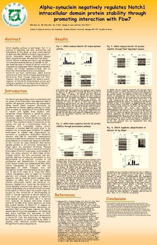
Fig. 1. SNCA reduces Notch1-IC transcriptional Fig. 3. SNCA reduces Notch1-IC protein
Notch signaling pathway is well known that it is activity. stability through Fbw7 dependent manner.
involved in regulating cell fate, proliferation and
homeostasis. In this study, we show a novel function
of alpha-synuclein (SNCA) to promote degradation of
Notch1 intracellular domain (Notch1-IC) through
Fbw7, ubiquitin E3 ligase. We identified that SNCA
inhibits Notch1 transcription activity and diminishes
the interaction between Notch1-IC and RBP-Jk. We
also found decrease of Notch1-IC protein stability by
exogenous and endogenous SNCA through proteasomal
pathway, not through lysosomal pathway. And, we
found that SNCA promotes interaction between
Notch1-IC and Fbw7. Furthermore, SNCA directly
interacts with Fbw7. SNCA increases ubiquitination of
Notch-IC by Fbw7 through interaction with Fbw7.
Together, these results suggest that SNCA is a novel
regulator of Notch1-IC transcriptional activity with
acting as an enhancer of the interaction of Notch1-IC
and Fbw7 with increasing degradation of Notch1-IC.
Introduction (A–B) HEK293 cells were transfected with expression vectors encoding (A) HEK293 cells were transiently transfected with expression vectors
encoding for shCon or shFbw7, Myc-Notch1-IC and Myc-SNCA. 48 h
for 4xCSL-Luc and -galatosidase, (A) along with Notch1-IC and 100 ng
(+), 200 ng (+ +), 300 ng (+ + +)of SNCA, (B) and control shRNA, or after transfection, the cells werelysates and lysates were immunoblotted
Notch1 shRNA, as indicated. (C) HEK293 cells were transiently with anti-Myc antibody. The expressions of Myc-SNCA or Fbw7 were
transfected with shCon or shSNCA as indicated. 48 h after transfection, analyzed via immunoblotting using anti-Myc and anti-Fbw7antibody,
thecells were lysed, and lysates were subjected to immunoprecipitation respectively. (B) HEK293 cells were transfected with expression vectors
The Notch signaling pathway, which is evolutionally well using IgG or anti-Notch1 antibodies, as indicated. The encoding for Myc-Notch1-IC, Myc-SNCA and Flag-Fbw7 or Fbw7 F. 48
con-versed, is well known that it has important roles to immunoprecipitates were immunoblotted withanti-RBP-Jk antibody. The h after transfection,the cells were lysates and lysates were
immunoblotted with anti-Myc antibody. The expressions of Myc-SNCA or
expressions of Notch1-IC, RBP-Jk or SNCA were analyzed via
regulate cell apoptosis, proliferation and homeostasis. immunoblotting using anti-Notch1, anti- RBP-Jk and anti-SNCA antibody, Flag-Fbw7 or Fbw7 F were analyzed via immunoblottingusing anti-Myc
Among several Notch receptors (Notch1–4), we respectively.(D) HEK293 cells were transfected with expression vectors and anti-Flag antibody, respectively. (C) HEK293 cells were transfected
with expression vectors encoding for Myc-SNCA and Flag-Fbw7. 48 h
encoding for Myc-Notch1-IC, Flag-RBP-Jk and Myc-SNCA as indicated.
concentrated on Notch1 signaling path-way [1]. Right 48 h after transfection, the cells lysates weresubjected to after transfection,the cells lysates were subjected to
after activation by interaction of extracellular domain immunoprecipitation analysis with anti-Flag. The immunoprecipitates were immunoprecipitation analysis with anti-Flag. The immunoprecipitates were
of Notch1 with ligands from neighbor cells, then immunoblotted with anti-Myc. The expressions of Myc-Notch1-IC, then immunoblotted with anti-Myc. The expressions of Myc-SNCA or
Flag-Fbw7 were analyzed via immunoblotting using anti-Myc and anti-Flag
Flag-RBP-Jkor Myc-SNCA were analyzed via immunoblotting using anti-
intracellular domain ofNotch1 is cleaved by gamma- Myc and anti-Flag antibody, respectively. Data are shown as mean ± antibody, respectively. (D) HEK293 cells lysates were subjected to
secretase complex, which results in release of SD(n = 3), *p < 0.05. immunoprecipitationusing IgG or anti-Fbw7 antibodies, as indicated. The
immunoprecipitates were immunoblotted with anti-SNCA antibody. The
intracellular domain of Notch1 (Notch1-IC) along with expressions of SNCA, Notch1 or Fbw7 wereanalyzed via immunoblotting
cleavage and release of extracellular domain of Notch1 using anti-SNCA, anti-Notch1 and anti-Fbw7 antibody, respectively. (A–
[3,5,22]. Then, released Notch1-IC translocate to the D) The data presented here are representative from three
independentexperiments.
nucleus, and acts as a transcriptional factor by Fig. 2. SNCA down-regulates Notch1-IC protein
interacting with recombining binding protein stability through proteasomal pathway.
Suppressor of Hairless (RBP-Jk), which binds to the Fig. 4. SNCA regulates ubiquitination of
promoter site of target genes of Notch1 to regulate Notch1-IC by Fbw7.
transcription by complex with transcriptional co-
repressors or co-activators [11,18].When Notch1-IC
interacts with RBP-Jk, associated transcriptional
repressors to RBP-Jk are detached, on the other hand,
transcriptional co-activators are associated with RBP-
Jk [23]. Overall, not along Notch1-IC; when Notch1
signaling pathway is not activated, transcription of
target genes are prevented [21,23,24]. Through
control of transcriptional factors, Notch1-IC activates
transcription of Notch1 target genes, as an example of
them, Hes (Hairy enhancer of split) family [12,14].
Although Notch1 has both pro-survival and pro-
apoptotic functions according to environment of each
tissues,Notch1 is an important activator of cell (A) HEK293 cells were transiently transfected with expression vectors
proliferation and differentiation in neuronal cells encoding for Myc- Notch1-IC and 200 ng (+), 400 ng (+ +), 600 ng (+ +
[4,20]. Recent studies have demonstrated that SNCA +) of Myc-SNCA, as indicated.48 h after transfection, the cells were
lysed and lysates were immunoblotted with anti-Myc antibody. The
inhibits neurogenesis of mouse stem cells and expressions of Myc-SNCA were analyzed via immunoblottingusing anti-
hippocampus of SNCA transgenic mice, and Myc antibody. (B) HEK293 cells were transfected for 48 h with
expression vectors encoding for control shRNA and SNCA shRNA, and
neurogenesis of adult rat hippocampus neural progeni- treated with 100 M DMSOor 100 M cycloheximide for the indicated
tors as well [7,8]. Crews et al. found that SNCA periods of time. The lysates were immunoblotted with anti-Notch1 (A) HEK293 cells were transiently transfected with shCon or shSNCA as
reduces neurogenesis which is caused by increased antibody. (C) HEK293 cells were transfected for 48 h withexpression indicated. 48 h after transfection, the cells were lysed, and lysates
vectors encoding for Flag-Notch1-IC and Myc-SNCA. HEK293 cells were
apoptosis and reduced proliferation of neuronal treated with 25 M of ALLN, 5 M of MG132 and 100 nM of Epoxomicin were subjected to immunopre-cipitation using IgG or anti-Fbw7
antibodies, as indicated. The immunoprecipitates were immunoblotted
progenitor cells by hindering Notch1 signaling; down- for 6 h. The lysateswere immunoblotted with anti-Flag antibody. The with anti-Notch1 antibody. The expressions of Notch1-IC, Fbw7or
expressions of Myc-SNCA were analyzed via immunoblotting using anti-
regulation of mRNA level of Hes-5, and protein Myc antibody. (D) HEK293 cells were transfectedfor 48 h with SNCA were analyzed via immunoblotting using anti-Notch1, anti-Fbw7
and anti-SNCA antibody, respectively. (B) HEK293 cells were
stability offull-length Notch1 and Notch1-IC [7]. expression vectors encoding for Myc-Notch1-IC and Myc-SNCA. transfected with expression vectorsencoding for Myc-tagged Notch1-IC,
Moreover, Desplats et al. found that the inhibition of HEK293 cells were treated with 100 M of Chloroquine and 50 mM of shFbw7, shSNCA and HA-tagged ubiquitin, as indicated. 48 h after
NH4Cl for 6 h. The lysateswere immunoblotted with anti-Myc antibody.
Notch1 signaling is derived from capacity of SNCA to The expressions of Myc-SNCA were analyzed via immunoblotting using transfection, the cells lysates were subjected to
immunoprecipitationanalysis with anti-Myc. The immunoprecipitates were
bind to the promoter site of Notch1 with p53 to hinder anti-Myc antibody. (A–D) The data presented here arerepresentative then immunoblotted with anti-HA. (C) HEK293 cells were transfected
from three independent experiments.
transcription of Notch1 [8].In the present study, we with expression vectors encoding for 4xCSL-Lucand -galatosidase, along
with Notch1-IC, SNCA and Fbw7 F, as indicated. Data are shown as
focused on that attenuation of Notch1signaling by mean ± SD(n = 3), *p ≤ 0.005; NS: non-significant.
SNCA is through regulation of Notch1 protein sta- References
bility. First, we confirmed decrease of Notch1-IC
transcriptional activity by SNCA, and it is derived Conclusions
from reduction of physical inter-action between 1. References[1] S. Artavanis-Tsakonas, M.D. Rand, R.J. Lake, Notch
Notch1-IC and RBP-Jk by SNCA. Moreover, we found signaling: cell fate controland signal integration in development, DiscussionIn this study, we provide the mechanism that how SNCA reg-
that protein stability of Notch1-IC is reduced by Science 284 (1999) 770–776.[2] S.H. Baek, M.Y. Kim, J.S. Mo, ulates Notch1 signaling pathway. We describe that SNCA playsan important
E.J. Ann, K.S. Lee, J.H. Park, J.Y. Kim, M.S. Seo, E.J.Choi, H.S.
SNCA through proteasomal pathway. Importantly, we Park, Zinc-induced downregulation of notch signaling is associatedwith role in regulation of Notch1 signaling pathway withacting as an enhancer of
the interaction of Notch1-IC and Fbw7with promoting degradation of
identified Fbw7 as an E3ligase required for SNCA- cytoplasmic retention of notch1-ic and rbp-jk via pi3k-akt Notch1-IC. The Notch1 signalingpathway regulates cell fate through cross-
signalingpathway, Cancer Lett. 255 (2007) 117–126.[3] C.M.
induced Notch1-IC protein degradation. We identified Blaumueller, H. Qi, P. Zagouras, S. Artavanis-Tsakonas, talks between neighbor-ing cells by affecting the processes of proliferation,
that SNCA facilitates the interaction of Fbw7 Intracellularcleavage of notch leads to a heterodimeric receptor on differentiationand apoptosis. Canonical Notch1 signaling requires that
Notch1-ICinteracts with the DNA-binding protein complex RBP-Jk. This
withNotch1-IC and thereby facilitates ubiquitination the plasma membrane,Cell 90 (1997) 281–291.[4] V. Bolos, J. com-plex is able to activate the transcription of various target genes suchas
Grego-Bessa, J.L. de la Pompa, Notch signaling in development
of Notch1-IC. From this observation, we implicated andcancer, Endocr. Rev. 28 (2007) 339–363.[5] C. Brou, F. Logeat, HES family [1].
that down-regulation of Notch1signaling by SNCA is N. Gupta, C. Bessia, O. LeBail, J.R. Doedens, A. Cumano, P.Roux,
R.A. Black, A. Israel, A novel proteolytic cleavage involved in
resulted from Notch1-IC protein destabilization notchsignaling: the role of the disintegrin-metalloprotease tace, Mol.
through Fbw7-mediated ubiquitination. Cell 5 (2000)207–216.[6] C. Cook, J. Gass, J. Dunmore, J. Tong, J.
Taylor, J. Eriksen, E. McGowan, J. Lewis,J. Johnston, L. Petrucelli,
Aging is not associated with proteasome impairmentin ups reporter
mice, PLoS One 4 (2009) e5888.[7] L. Crews, H. Mizuno, P.
Desplats, E. Rockenstein, A. Adame, C. Patrick, B.Winner, J.
Winkler, E. Masliah, Alpha-synuclein alters notch-1 expression
andneurogenesis in mouse embryonic stem cells and in the hippocampus
oftransgenic mice, J. Neurosci.: Off. J. Soc. Neurosc. 28 (2008)
4250–4260.[8] P. Desplats, B. Spencer, L. Crews, P. Pathel, D.
Morvinski-Friedmann, K.Kosberg, S. Roberts, C. Patrick, B. Winner,
J. Winkler, E. Masliah,Alpha-synuclein induces alterations in adult
neurogenesis in parkinsondisease models via p53-mediated repression
of notch1, J. Biol. Chem. 287(2012) 31691–31702.

