


Page 19 - ebook
P. 19
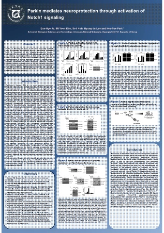
Parkin mediates neuroprotection through activation of

                     Notch1 signaling


                              Eun-Hye Jo, Mi-Yeon Kim, So-I Noh, Hyung-Ju Lee and Hee-Sae Park *
                     School of Biological Sciences and Technology, Chonnam National University, Gwangju 500-757, Republic of Korea


                  Abstract                 Figure 1. Parkin activates Notch1-IC   Figure 4. Parkin reduces neuronal apoptosis
                                           transcriptional activity.             through the Notch1 signaling pathway.
     Parkin, an E3 ubiquitin ligase, is the most frequently mutated
     gene in hereditary Parkinson’s disease. Inactivation of Parkin
     leads to impairment of the ubiquitin–proteasome system,
     resulting in the accumulation of misfolded or aggregated proteins
     and ensuing neurodegeneration. In this study, we show that
     Parkin positively regulates the Notch1 signaling pathway.
     Overexpression of Parkin stabilized Notch1-IC protein levels,
     whereas knockdown of Parkin decreased Notch1-IC protein
     stability. Notably, overexpression of Parkin disrupted oxidative
     stress-induced apoptosis in neuronal cells. However, knockdown
     of Notch1 inhibited Parkin-induced neuronal cell survival.
     Together, these results indicate that Parkin is a novel regulator of
     the Notch1 signaling pathway,                                                (a) Intracellular reactive oxygen species (ROS) generation was
                                                                                  analyzed by staining with 10 μM DCFHDA for 15 min in SH-SY5Y
                                                                                  cells transfected with His-Parkin and shNotch1-IC and treated
                                                                                  with 2 mM H2O2 for 20 min as indicated. (b) Apoptosis assay of
                                                                                  the SH-SY5Y cells transfected with His-Parkin and shNotch1-IC
                                           (a–c) Luciferase assay of HEK293 cells transiently transfected  and treated with 2 mM H2O2 for 20 min Apoptotic cells were
                 Introduction              with plasmids expressing Parkin and Notch1-IC. Transcriptional  stained with 5 μl annexin V-FITC for 5 min in the dark. Results of
                                           activity of Notch1-IC was measured using luciferase reporter  three independent experiments are presented as mean ±SD. (c) A
                                           plasmids, (a) 4xCSL-Luc, (b) Hes1-Luc, and (c) Hes5-Luc. (d)
     Parkinson’s disease (PD) is the most common movement  Transcriptional activity of Notch1-IC in HEK293 cells co-  working model of Notch1 activation by Parkin. Parkin interacts
     disorder in humans and is characterized by the loss of dopamine-             with Fbw7 and induces its ubiquitination and degradation.
     producing neurons in the substantia nigra, a region in the  transfected with Notch1-IC and either Parkin WT or R42P PD  Degradation of Fbw7 stabilizes Notch1-IC and stimulates the
                                           pathogenic mutant. (e) Luciferase assay of HEK293 cells
     midbrain, which is implicated in motor control. Among the genes  transiently transfected with plasmids expressing Notch1-IC and  Notch1 signaling pathway, thus accelerating neuronal survival.
     that are responsible for monogenic familial variants of PD, Parkin
     appears to play a prominent role, accounting for the majority of  Parkin shRNA. (f) Western blot analysis of Parkin using lysates of
                                           HEK293 cells dose dependently transfected with shParkin.
     autosomal recessive PD cases. Parkin is an E3 ubiquitin protein
     ligase targeting substrate proteins for proteasomal degradation.  Results of three independent experiments are presented as mean
                                           ±SD.
     Mutations in Parkin, which disrupt its catalytic activity, lead to the
     accumulation of toxic substrates that damage dopaminergic                    Figure 5. Parkin significantly stimulates
     neurons, consequently causing autosomal recessive PD[1].                     neuronal protection under oxidative stress by a
     Parkin is transcriptionally upregulated under various stress
     conditions including mitochondrial and endoplasmic reticulum  Figure 2. Parkin stimulates the interaction   Notch1-mediated pathway.
     stress,  proteotoxic  stress,  excitotoxicity,  proapoptotic  between Notch1-IC and RBP-Jκ.
     stimulation, as well as overexpression of toxic, misfolded protein
     species, and shows wide neuroprotective activity in cellular and
     animal  models[2–4].  Parkin  promotes  neuroprotection  by
     regulating  various  biological  mechanisms,  including
     mitochondrial  integrity,  proteasomal  degradation  of  toxic
     substances, and nondegradative ubiquitin signaling within cell
     death or viability pathways [5]. Parkin is responsible for proper
     mitochondrial  function  and  promotes  autophagosomal
     engulfment of damaged mitochondria in a process termed
     mitophagy. Upon mitochondrial depolarization, Parkin cooperates               (c) A working model of Notch1 activation by Parkin. Parkin
     with mitochondrial PTEN-induced putative kinase 1 (PINK1).                    interacts with Fbw7 and induces its ubiquitination and
     PINK1 expression was found to be necessary for the recruitment                degradation. Degradation of Fbw7 stabilizes Notch1-IC and
     of Parkin to depolarized mitochondria. Under stress conditions,               stimulates the Notch1 signaling pathway, thus accelerating
     Parkin stimulates mitochondrial biogenesis and cell survival of               neuronal survival.
     dopaminergic neurons through ubiquitination of its target  (a) Co-IP of Notch1-IC and RBP-Jκ in HEK293 cells transiently
     proteins. Furthermore, Parkin can also influence other cell death  transfected with Myc-Notch1-IC, Flag-RBP-Jκ, and His-Parkin
     and viability pathways through a nondegradative mechanism by  expression vectors as indicated. Myc-Notch1-IC, Flag-RBP-Jκ,
     modulating ubiquitin signaling in the nuclear factor kappa B (NF-  and  His-Parkin  expression  was  analyzed  using  specific
     κB)  pathway,  activation  of  c-Jun  N-terminal  kinase,  p53  antibodies, respectively. (b) Endogenous co-IP of Notch1-IC and
     expression, and mitochondrial translocation of Bax [6–8].  RBP-Jκ in HEK293 cells transiently transfected with shControl
     Abnormal activation of the Notch1 pathway is associated with  and shParkin. Notch1-IC, RBP-Jκ, and Parkin expressions were
     several  neurodegenerative  diseases  including  amyotrophic  analyzed  using  anti-Notch1,  anti-RBP-Jκ,  and  anti-Parkin  Conclusion
     lateral sclerosis, Down’s syndrome, Alzheimer’s, and Pick’s  antibodies,  respectively.  Results  of  three  independent
     disease. Notch1 promotes cell survival in the developing nervous  experiments are presented as mean ±SD.
     system . Although the Notch1 signaling pathway is known to be                Previously, it was shown that the Notch1 signaling pathway
     important                                                                    is important for neural stem cell maintenance and proper
     during neuronal degeneration, its regulatory mechanism remains               neurogenesis in the developing mammalian nervous
     to be determined. In this study, we identified Parkin as an  Figure 3. Parkin induces Notch1-IC protein   system and in the adult brain. Importantly, Notch1 also
     activator of Notch1 signaling, indicating that Parkin mediates its           plays a role in neuronal differentiation and neuronal
     neuroprotective effect by modulating Fbw7-mediated protein  stability in an Fbw7-dependent manner.   survival. These data indicate that Notch1 is required not
     degradation of Notch1.                                                       only for the formation but also for the maintenance of
                                                                                  neurons. In the present study, we focused on Parkin-
                                                                                  mediated activation of Notch1 signaling, leading to
                                                                                  neuronal survival. Loss of Parkin E3 ligase activity leads to
                 References                                                       impairment of the ubiquitin–proteasome system, resulting
                                                                                  in the accumulation of misfolded proteins and ensuing
                                                                                  neurodegeneration. Parkin ubiquitinated and degraded the
     1 Dawson TM, Dawson VL. The role of parkin in familial and
      sporadic                                                                    target proteins of numerous signaling pathways. Parkin
     Parkinson’s disease. Mov Disord 2010; 25 (Suppl 1):S32–S39.                  ubiquitinates the proapoptotic factor Bax both in vivo and
     2 Yang YX, Muqit MM, Latchman DS. Induction of parkin                        in vitro. Indeed, Parkin also ubiquitinated NF-κB essential
      expression in the                                                           modulator resulting in protection from stress-induced cell
     presence of oxidative stress. Eur J Neurosci 2006; 24:1366–1372.             death. Here, we provided the correlation between Parkin
     3 LaVoie MJ, Cortese GP, Ostaszewski BL, Schlossmacher MG.                   and the Notch1 signaling pathway (Fig. 4c). Notch1-IC
      The effects of oxidative stress on parkin and other E3 ligases.             protein stability is regulated by ubiquitination. Fbw7, an F-
      J Neurochem 2007; 103:2354–2368.                                            box protein, is known to be a nuclear ubiquitin E3 ligase
     4 Wong ES, Tan JM, Wang C, Zhang Z, Tay SP, Zaiden N, et al.                 and  regulates  Notch1-IC  proteasome-dependent
      Relative sensitivity of parkin and other cysteine-containing                degradation. We confirmed that Fbw7 is necessary for
      enzymes to stress-induced solubility alterations. J Biol Chem
      2007; 282:12310–12318.               (a)Western blot analysis using antibodies against Flag and Myc in lysates of  Parkinmediated regulation of Notch1-IC protein stability.
     5 Winklhofer KF. Parkin and mitochondrial quality control: toward   HEK293 cells dose-dependently expressing Flag-Notch1-IC and Myc-Parkin. (b)  Parkin inhibited the interaction between Notch1-IC and
                                           Cycloheximide (CHX) chase assay for the half-life of Notch1-IC of SH-SY5Y
      assembling the puzzle. Trends Cell Biol 2014; 24:332–341.  cells transfected with shControl and shParkin. Cells were treated with either  Fbw7, resulting in reduced ubiquitination of Notch1-IC.
     6 da Costa CA, Sunyach C, Giaime E, West A, Corti O, Brice A, et   100 μM DMSO or 100 μM CHX for the indicated times. The lysates were  These findings reveal a novel mechanism for Parkin-
      al. Transcriptional repression of p53 by parkin and impairment   immunoblotted using an anti-Notch1-IC and anti-β-actin antibody. (c) Western  mediated regulation of the Notch1 signaling pathway.
      by mutations associated with autosomal recessive juvenile   blot analysis, using antibodies against Flag and Myc, of lysates from HEK293
      Parkinson’s disease. Nat Cell Biol 2009; 11:1370–1375.  cells dose-dependently expressing Flag-Fbw7 and Myc-Parkin. (d)Westernblot
                                           analysis of Myc-Notch1-IC of HEK293 cells cotransfected with Myc-Notch1-IC,
     7 Cha GH, Kim S, Park J, Lee E, Kim M, Lee SB, et al. Parkin  Flag-Fbw7, and Myc-Parkin. (e) Co-IP of Notch1-IC and RBP-Jκ in HEK293 cells
      negatively regulates JNK pathway in the dopaminergic neurons   transiently transfected with Myc-Notch1-IC, Flag-Fbw7, and His-Parkin
      of Drosophila. Proc Natl Acad Sci USA 2005; 102:10345–10350.  expression vectors as indicated. (f) Endogenous co-IP of Notch1-IC and Fbw7
     8 Henn IH, Bouman L, Schlehe JS, Schlierf A, Schramm JE,   in HEK293 cells transiently transfected with shControl and shParkin. (g)
      Wegener E, et al. Parkin mediates neuroprotection through   Luciferase assay of HEK293 cells transiently transfected with plasmids
                                           expressing Myc-Notch1-IC, Fbw7 shRNA, and Parkin shRNA as indicated.
      activation of IkappaB kinase/nuclear factor-kappaB signaling. J   Results of three independent experiments are presented as mean ±SD.
      Neurosci 2007; 27:1868–1878.

